3ZI5

Crystal STRUCTURE OF RESTRICTION ENDONUCLEASE BFII C-TERMINAL RECOGNITION DOMAIN IN COMPLEX WITH COGNATE DNA


Experimental Data Snapshot

  • Method: X-RAY DIFFRACTION
  • Resolution: 3.20 Å
  • R-Value Free: 0.226 
  • R-Value Work: 0.176 
  • R-Value Observed: 0.180 

wwPDB Validation   3D Report Full Report


This is version 1.4 of the entry. See complete history


Literature

Structural Insight Into the Specificity of the B3 DNA-Binding Domains Provided by the Co-Crystal Structure of the C-Terminal Fragment of Bfii Restriction Enzyme

Golovenko, D.Grazulis, S.Manakova, E.Sasnauskas, G.Siksnys, V.Zakrys, L.Zaremba, M.

(2014) Nucleic Acids Res 42: 4113

  • DOI: https://doi.org/10.1093/nar/gkt1368
  • Primary Citation of Related Structures:  
    3ZI5

  • PubMed Abstract: 

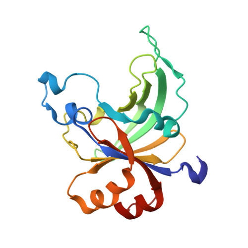
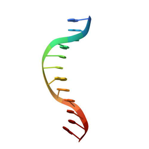
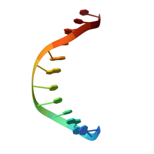
    The B3 DNA-binding domains (DBDs) of plant transcription factors (TF) and DBDs of EcoRII and BfiI restriction endonucleases (EcoRII-N and BfiI-C) share a common structural fold, classified as the DNA-binding pseudobarrel. The B3 DBDs in the plant TFs recognize a diverse set of target sequences. The only available co-crystal structure of the B3-like DBD is that of EcoRII-N (recognition sequence 5'-CCTGG-3'). In order to understand the structural and molecular mechanisms of specificity of B3 DBDs, we have solved the crystal structure of BfiI-C (recognition sequence 5'-ACTGGG-3') complexed with 12-bp cognate oligoduplex. Structural comparison of BfiI-C-DNA and EcoRII-N-DNA complexes reveals a conserved DNA-binding mode and a conserved pattern of interactions with the phosphodiester backbone. The determinants of the target specificity are located in the loops that emanate from the conserved structural core. The BfiI-C-DNA structure presented here expands a range of templates for modeling of the DNA-bound complexes of the B3 family of plant TFs.


  • Organizational Affiliation

    Department of Protein-DNA Interactions, Institute of Biotechnology, Vilnius University, Graičiūno 8, LT-02241, Vilnius, Lithuania.


Macromolecules

Find similar proteins by:  (by identity cutoff)  |  3D Structure
Entity ID: 1
MoleculeChains Sequence LengthOrganismDetailsImage
RESTRICTION ENDONUCLEASE
A, D
166Cytobacillus firmusMutation(s): 0 
EC: 3.1.21.4
UniProt
Find proteins for Q9F4C9 (Cytobacillus firmus)
Explore Q9F4C9 
Go to UniProtKB:  Q9F4C9
Entity Groups  
Sequence Clusters30% Identity50% Identity70% Identity90% Identity95% Identity100% Identity
UniProt GroupQ9F4C9
Sequence Annotations
Expand
  • Reference Sequence

Find similar nucleic acids by:  Sequence   |   3D Structure  

Entity ID: 2
MoleculeChains LengthOrganismImage
5'-D(*AP*GP*CP*AP*CP*TP*GP*GP*GP*TP*CP*GP)-3'
B, E
12synthetic construct
Sequence Annotations
Expand
  • Reference Sequence

Find similar nucleic acids by:  Sequence   |   3D Structure  

Entity ID: 3
MoleculeChains LengthOrganismImage
5'-D(*CP*GP*AP*CP*CP*CP*AP*GP*TP*GP*CP*TP)-3'
C, F
12synthetic construct
Sequence Annotations
Expand
  • Reference Sequence
Experimental Data & Validation

Experimental Data

  • Method: X-RAY DIFFRACTION
  • Resolution: 3.20 Å
  • R-Value Free: 0.226 
  • R-Value Work: 0.176 
  • R-Value Observed: 0.180 
  • Space Group: P 65
Unit Cell:
Length ( Å )Angle ( ˚ )
a = 175.18α = 90
b = 175.18β = 90
c = 35.789γ = 120
Software Package:
Software NamePurpose
PHENIXrefinement
MOSFLMdata reduction
TRUNCATEdata scaling
MOLREPphasing

Structure Validation

View Full Validation Report


Entry History 

Deposition Data

Revision History  (Full details and data files)

  • Version 1.0: 2014-01-15
    Type: Initial release
  • Version 1.1: 2014-01-29
    Changes: Database references
  • Version 1.2: 2014-04-16
    Changes: Database references
  • Version 1.3: 2019-05-08
    Changes: Advisory, Data collection, Experimental preparation, Other
  • Version 1.4: 2023-12-20
    Changes: Advisory, Data collection, Database references, Other, Refinement description