3ZFH | pdb_00003zfh

Crystal structure of Pseudomonas aeruginosa inosine 5'-monophosphate dehydrogenase


Experimental Data Snapshot

  • Method: X-RAY DIFFRACTION
  • Resolution: 2.25 Å
  • R-Value Free: 
    0.190 (Depositor), 0.171 (DCC) 
  • R-Value Work: 
    0.148 (Depositor) 
  • R-Value Observed: 
    0.150 (Depositor) 

Starting Model: experimental
View more details

wwPDB Validation 3D Report Full Report

Validation slider image for 3ZFH

This is version 1.3 of the entry. See complete history

Literature

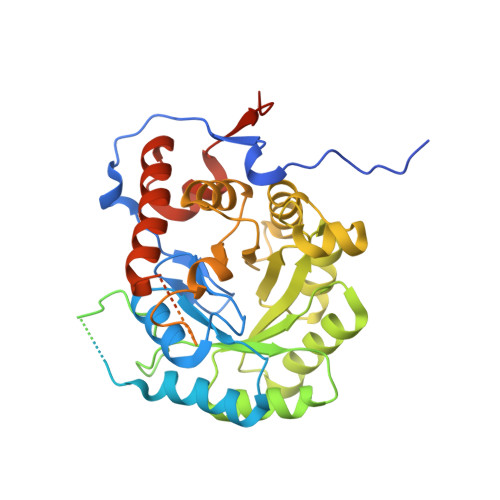
Structure of Pseudomonas Aeruginosa Inosine 5'-Monophosphate Dehydrogenase

Rao, V.A.Shepherd, S.M.Owen, R.Hunter, W.N.

(2013) Acta Crystallogr Sect F Struct Biol Cryst Commun 69: 243

  • DOI: https://doi.org/10.1107/S1744309113002352
  • Primary Citation Related Structures: 
    3ZFH

  • PubMed Abstract: 

    Inosine 5'-monophosphate dehydrogenase (IMPDH) represents a potential antimicrobial drug target. The crystal structure of recombinant Pseudomonas aeruginosa IMPDH has been determined to a resolution of 2.25 Å. The structure is a homotetramer of subunits dominated by a (β/α)8-barrel fold, consistent with other known structures of IMPDH. Also in common with previous work, the cystathionine β-synthase domains, residues 92-204, are not present in the model owing to disorder. However, unlike the majority of available structures, clearly defined electron density exists for a loop that creates part of the active site. This loop, composed of residues 297-315, links α8 and β9 and carries the catalytic Cys304. P. aeruginosa IMPDH shares a high level of sequence identity with bacterial and protozoan homologues, with residues involved in binding substrate and the NAD+ cofactor being conserved. Specific differences that have been proven to contribute to selectivity against the human enzyme in a study of Cryptosporidium parvum IMPDH are also conserved, highlighting the potential value of IMPDH as a drug target.


  • Organizational Affiliation
    • Division of Biological Chemistry and Drug Discovery, College of Life Sciences, University of Dundee, Dow Street, Dundee DD1 5EH, Scotland.

Macromolecule Content 

  • Total Structure Weight: 54.22 kDa 
  • Atom Count: 2,298 
  • Modeled Residue Count: 299 
  • Deposited Residue Count: 510 
  • Unique protein chains: 1

Macromolecules

Find similar proteins by:|  3D Structure
Entity ID: 1
MoleculeChains  Sequence LengthOrganismDetailsImage
INOSINE 5'-MONOPHOSPHATE DEHYDROGENASE510Pseudomonas aeruginosa PAO1Mutation(s): 0 
EC: 1.1.1.205
UniProt
Find proteins for Q9HXM5 (Pseudomonas aeruginosa (strain ATCC 15692 / DSM 22644 / CIP 104116 / JCM 14847 / LMG 12228 / 1C / PRS 101 / PAO1))
Explore Q9HXM5 
Go to UniProtKB:  Q9HXM5
Entity Groups
Sequence Clusters30% Identity50% Identity70% Identity90% Identity95% Identity100% Identity
UniProt GroupQ9HXM5
Sequence Annotations
Expand
Reference Sequence

Small Molecules

Ligands 1 Unique
IDChains Name / Formula / InChI Key2D Diagram3D Interactions
CL

Query on CL


Download:Ideal Coordinates CCD File
B [auth A]CHLORIDE ION
Cl
VEXZGXHMUGYJMC-UHFFFAOYSA-M

Experimental Data & Validation

Experimental Data

  • Method: X-RAY DIFFRACTION
  • Resolution: 2.25 Å
  • R-Value Free:  0.190 (Depositor), 0.171 (DCC) 
  • R-Value Work:  0.148 (Depositor) 
  • R-Value Observed: 0.150 (Depositor) 
Space Group: I 4
Unit Cell:
Length ( Å )Angle ( ˚ )
a = 115.546α = 90
b = 115.546β = 90
c = 56.443γ = 90
Software Package:
Software NamePurpose
REFMACrefinement
XDSdata reduction
SCALAdata scaling
PHASERphasing

Structure Validation

View Full Validation Report


Entry History 

Deposition Data

Revision History  (Full details and data files)

  • Version 1.0: 2013-01-16
    Type: Initial release
  • Version 1.1: 2013-03-13
    Changes: Database references
  • Version 1.2: 2013-04-17
    Changes: Database references
  • Version 1.3: 2023-12-20
    Changes: Data collection, Database references, Other, Refinement description