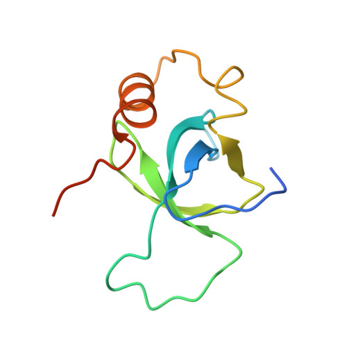
Nucleosomal DNA Binding Drives the Recognition of H3K36-Methylated Nucleosomes by the Psip1-Pwwp Domain.
Van Nuland, R., Van Schaik, F.M., Simonis, M., Van Heesch, S., Cuppen, E., Boelens, R., Timmers, H.T.M., Van Ingen, H.(2013) Epigenetics Chromatin 6: 12
- PubMed: 23656834 Search on PubMedSearch on PubMed Central
- DOI: https://doi.org/10.1186/1756-8935-6-12
- Primary Citation Related Structures:
3ZEH - PubMed Abstract:
Recognition of histone modifications by specialized protein domains is a key step in the regulation of DNA-mediated processes like gene transcription. The structural basis of these interactions is usually studied using histone peptide models, neglecting the nucleosomal context. Here, we provide the structural and thermodynamic basis for the recognition of H3K36-methylated (H3K36me) nucleosomes by the PSIP1-PWWP domain, based on extensive mutational analysis, advanced nuclear magnetic resonance (NMR), and computational approaches. The PSIP1-PWWP domain binds H3K36me3 peptide and DNA with low affinity, through distinct, adjacent binding surfaces. PWWP binding to H3K36me nucleosomes is enhanced approximately 10,000-fold compared to a methylated peptide. Based on mutational analyses and NMR data, we derive a structure of the complex showing that the PWWP domain is bound to H3K36me nucleosomes through simultaneous interactions with both methylated histone tail and nucleosomal DNA. Concerted binding to the methylated histone tail and nucleosomal DNA underlies the high- affinity, specific recognition of H3K36me nucleosomes by the PSIP1-PWWP domain. We propose that this bipartite binding mechanism is a distinctive and general property in the recognition of histone modifications close to the nucleosome core.
- NMR Spectroscopy Research Group, Bijvoet Center for Biomolecular Research, Utrecht University Utrecht, Padualaan 8, Utrecht, CH, 3854, The Netherlands. h.vaningen@uu.nl.
Organizational Affiliation: