3ZDV | pdb_00003zdv

Crystal structure of the LecB lectin from Pseudomonas aeruginosa in complex with Methyl 6-(2,4,6-trimethylphenylsulfonylamido)-6-deoxy-alpha-D-mannopyranoside


Experimental Data Snapshot

  • Method: X-RAY DIFFRACTION
  • Resolution: 1.41 Å
  • R-Value Free: 
    0.145 (Depositor), 0.151 (DCC) 
  • R-Value Work: 
    0.115 (Depositor), 0.126 (DCC) 
  • R-Value Observed: 
    0.116 (Depositor) 

Starting Model: experimental
View more details

wwPDB Validation 3D Report Full Report

Validation slider image for 3ZDV

Ligand Structure Quality Assessment 


This is version 1.4 of the entry. See complete history

Literature

Discovery of Two Classes of Potent Glycomimetic Inhibitors of Pseudomonas Aeruginosa Lecb with Distinct Binding Modes.

Hauck, D.Joachim, I.Frommeyer, B.Varrot, A.Philipp, B.Moller, H.M.Imberty, A.Exner, T.E.Titz, A.

(2013) ACS Chem Biol 8: 1775

  • DOI: https://doi.org/10.1021/cb400371r
  • Primary Citation Related Structures: 
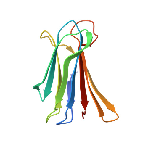
    3ZDV

  • PubMed Abstract: 

    The treatment of infections due to the opportunistic pathogen Pseudomonas aeruginosa is often difficult, as a consequence of bacterial biofilm formation. Such a protective environment shields the bacterium from host defense and antibiotic treatment and secures its survival. One crucial factor for maintenance of the biofilm architecture is the carbohydrate-binding lectin LecB. Here, we report the identification of potent mannose-based LecB inhibitors from a screening of four series of mannosides in a novel competitive binding assay for LecB. Cinnamide and sulfonamide derivatives are inhibitors of bacterial adhesion with up to a 20-fold increase in affinity to LecB compared to the natural ligand methyl mannoside. Because many lectins of the host require terminal saccharides (e.g., fucosides), such capped structures as reported here may offer a beneficial selectivity profile for the pathogenic lectin. Both classes of compounds show distinct binding modes at the protein, offering the advantage of a simultaneous development of two new lead structures as anti-pseudomonadal drugs with an anti-virulence mode of action.


  • Organizational Affiliation
    • Department of Chemistry, University of Konstanz, D-78457 Konstanz, Germany.

Macromolecule Content 

  • Total Structure Weight: 49.61 kDa 
  • Atom Count: 4,025 
  • Modeled Residue Count: 456 
  • Deposited Residue Count: 460 
  • Unique protein chains: 1

Macromolecules

Find similar proteins by:|  3D Structure
Entity ID: 1
MoleculeChains  Sequence LengthOrganismDetailsImage
FUCOSE-BINDING LECTIN PA-IIL
A, B, C, D
115Pseudomonas aeruginosa PAO1Mutation(s): 0 
UniProt
Find proteins for Q9HYN5 (Pseudomonas aeruginosa (strain ATCC 15692 / DSM 22644 / CIP 104116 / JCM 14847 / LMG 12228 / 1C / PRS 101 / PAO1))
Explore Q9HYN5 
Go to UniProtKB:  Q9HYN5
Entity Groups
Sequence Clusters30% Identity50% Identity70% Identity90% Identity95% Identity100% Identity
UniProt GroupQ9HYN5
Sequence Annotations
Expand
Reference Sequence

Small Molecules

Ligands 6 Unique
IDChains Name / Formula / InChI Key2D Diagram3D Interactions
F1A

Query on F1A


Download:Ideal Coordinates CCD File
H [auth A],
L [auth B],
R [auth C],
V [auth D]
2,4,6-trimethylbenzenesulfonamide
C9 H13 N O2 S
YECJUZIGFPJWGQ-UHFFFAOYSA-N
MMA

Query on MMA


Download:Ideal Coordinates CCD File
G [auth A],
K [auth B],
Q [auth C],
U [auth D]
methyl alpha-D-mannopyranoside
C7 H14 O6
HOVAGTYPODGVJG-VEIUFWFVSA-N
SO4

Query on SO4


Download:Ideal Coordinates CCD File
W [auth D]SULFATE ION
O4 S
QAOWNCQODCNURD-UHFFFAOYSA-L
GOL

Query on GOL


Download:Ideal Coordinates CCD File
M [auth B]GLYCEROL
C3 H8 O3
PEDCQBHIVMGVHV-UHFFFAOYSA-N
EDO

Query on EDO


Download:Ideal Coordinates CCD File
N [auth B]1,2-ETHANEDIOL
C2 H6 O2
LYCAIKOWRPUZTN-UHFFFAOYSA-N
CA

Query on CA


Download:Ideal Coordinates CCD File
E [auth A]
F [auth A]
I [auth B]
J [auth B]
O [auth C]
E [auth A],
F [auth A],
I [auth B],
J [auth B],
O [auth C],
P [auth C],
S [auth D],
T [auth D]
CALCIUM ION
Ca
BHPQYMZQTOCNFJ-UHFFFAOYSA-N

Experimental Data & Validation

Experimental Data

  • Method: X-RAY DIFFRACTION
  • Resolution: 1.41 Å
  • R-Value Free:  0.145 (Depositor), 0.151 (DCC) 
  • R-Value Work:  0.115 (Depositor), 0.126 (DCC) 
  • R-Value Observed: 0.116 (Depositor) 
Space Group: P 21 21 21
Unit Cell:
Length ( Å )Angle ( ˚ )
a = 50.53α = 90
b = 76.98β = 90
c = 99.66γ = 90
Software Package:
Software NamePurpose
REFMACrefinement
XDSdata reduction
XSCALEdata scaling
PHASERphasing

Structure Validation

View Full Validation Report


Ligand Structure Quality Assessment 


Entry History 

Deposition Data

Revision History  (Full details and data files)

  • Version 1.0: 2013-09-18
    Type: Initial release
  • Version 1.1: 2013-11-13
    Changes: Other
  • Version 1.2: 2017-09-13
    Changes: Data collection, Structure summary
  • Version 1.3: 2020-07-29
    Type: Remediation
    Reason: Carbohydrate remediation
    Changes: Data collection, Derived calculations, Other, Structure summary
  • Version 1.4: 2023-12-20
    Changes: Data collection, Database references, Refinement description, Structure summary