3ZCZ | pdb_00003zcz

Crystal structure of a complex between Actinomadura R39 DD-peptidase and a trifluoroketone inhibitor


Experimental Data Snapshot

  • Method: X-RAY DIFFRACTION
  • Resolution: 2.60 Å
  • R-Value Free: 
    0.237 (Depositor), 0.231 (DCC) 
  • R-Value Work: 
    0.193 (Depositor), 0.191 (DCC) 
  • R-Value Observed: 
    0.195 (Depositor) 

Starting Model: experimental
View more details

wwPDB Validation 3D Report Full Report

Validation slider image for 3ZCZ

Ligand Structure Quality Assessment 


This is version 1.3 of the entry. See complete history

Literature

Inhibition of Dd-Peptidases by a Specific Trifluoroketone: Crystal Structure of a Complex with the Actinomadura R39 Dd-Peptidase.

Dzhekieva, L.Adediran, S.A.Herman, R.Kerff, F.Duez, C.Charlier, P.Sauvage, E.Pratt, R.F.

(2013) Biochemistry 52: 2128

  • DOI: https://doi.org/10.1021/bi400048s
  • Primary Citation Related Structures: 
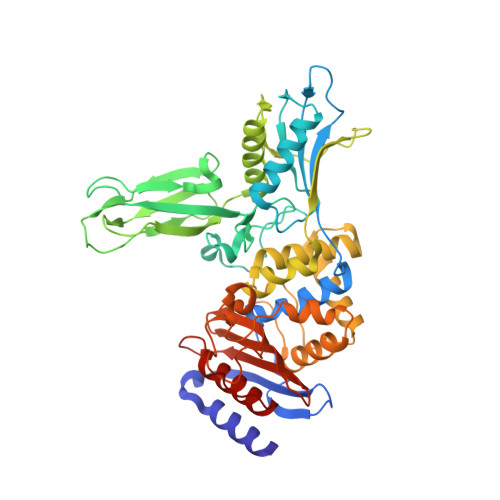
    3ZCZ

  • PubMed Abstract: 

    Inhibitors of bacterial DD-peptidases represent potential antibiotics. In the search for alternatives to β-lactams, we have investigated a series of compounds designed to generate transition state analogue structures upon reaction with DD-peptidases. The compounds contain a combination of a peptidoglycan-mimetic specificity handle and a warhead capable of delivering a tetrahedral anion to the enzyme active site. The latter includes a boronic acid, two alcohols, an aldehyde, and a trifluoroketone. The compounds were tested against two low-molecular mass class C DD-peptidases. As expected from previous observations, the boronic acid was a potent inhibitor, but rather unexpectedly from precedent, the trifluoroketone [D-α-aminopimelyl(1,1,1-trifluoro-3-amino)butan-2-one] was also very effective. Taking into account competing hydration, we found the trifluoroketone was the strongest inhibitor of the Actinomadura R39 DD-peptidase, with a subnanomolar (free ketone) inhibition constant. A crystal structure of the complex between the trifluoroketone and the R39 enzyme showed that a tetrahedral adduct had indeed formed with the active site serine nucleophile. The trifluoroketone moiety, therefore, should be considered along with boronic acids and phosphonates as a warhead that can be incorporated into new and effective DD-peptidase inhibitors and therefore, perhaps, antibiotics.


  • Organizational Affiliation
    • Department of Chemistry, Wesleyan University , Lawn Avenue, Middletown, Connecticut 06459, United States.

Macromolecule Content 

  • Total Structure Weight: 193.84 kDa 
  • Atom Count: 13,891 
  • Modeled Residue Count: 1,866 
  • Deposited Residue Count: 1,868 
  • Unique protein chains: 1

Macromolecules

Find similar proteins by:|  3D Structure
Entity ID: 1
MoleculeChains  Sequence LengthOrganismDetailsImage
D-ALANYL-D-ALANINE CARBOXYPEPTIDASE
A, B, C, D
467Actinomadura sp. R39Mutation(s): 0 
EC: 3.4.16.4
UniProt
Find proteins for P39045 (Actinomadura sp. (strain R39))
Explore P39045 
Go to UniProtKB:  P39045
Entity Groups
Sequence Clusters30% Identity50% Identity70% Identity90% Identity95% Identity100% Identity
UniProt GroupP39045
Sequence Annotations
Expand
Reference Sequence

Small Molecules

Ligands 3 Unique
IDChains Name / Formula / InChI Key2D Diagram3D Interactions
TFR

Query on TFR


Download:Ideal Coordinates CCD File
E [auth A],
M [auth B],
R [auth C],
X [auth D]
(2R)-2-amino-7-oxo-7-{[(2R,3S)-4,4,4-trifluoro-3-hydroxybutan-2-yl]amino}heptanoic acid
C11 H19 F3 N2 O4
GCYIIHHJPYOBBJ-BHNWBGBOSA-N
SO4

Query on SO4


Download:Ideal Coordinates CCD File
AA [auth D]
BA [auth D]
F [auth A]
G [auth A]
H [auth A]
AA [auth D],
BA [auth D],
F [auth A],
G [auth A],
H [auth A],
I [auth A],
L [auth A],
N [auth B],
O [auth B],
P [auth B],
Q [auth B],
S [auth C],
T [auth C],
U [auth C],
V [auth C],
W [auth C],
Y [auth D],
Z [auth D]
SULFATE ION
O4 S
QAOWNCQODCNURD-UHFFFAOYSA-L
MG

Query on MG


Download:Ideal Coordinates CCD File
CA [auth D],
DA [auth D],
J [auth A],
K [auth A]
MAGNESIUM ION
Mg
JLVVSXFLKOJNIY-UHFFFAOYSA-N

Experimental Data & Validation

Experimental Data

  • Method: X-RAY DIFFRACTION
  • Resolution: 2.60 Å
  • R-Value Free:  0.237 (Depositor), 0.231 (DCC) 
  • R-Value Work:  0.193 (Depositor), 0.191 (DCC) 
  • R-Value Observed: 0.195 (Depositor) 
Space Group: P 1 21 1
Unit Cell:
Length ( Å )Angle ( ˚ )
a = 103.653α = 90
b = 91.727β = 94.3
c = 106.882γ = 90
Software Package:
Software NamePurpose
REFMACrefinement
XDSdata reduction
XSCALEdata scaling

Structure Validation

View Full Validation Report


Ligand Structure Quality Assessment 


Entry History 

Deposition Data

Revision History  (Full details and data files)

  • Version 1.0: 2013-03-27
    Type: Initial release
  • Version 1.1: 2013-08-28
    Changes: Database references
  • Version 1.2: 2023-12-20
    Changes: Data collection, Database references, Derived calculations, Other, Refinement description
  • Version 1.3: 2024-11-13
    Changes: Structure summary