Crystal Structures of Murine Angiogenin-2 and -3 - Probing 'Structure - Function' Relationships Amongst Angiogenin Homologues.
Iyer, S., Holloway, D.E., Acharya, K.R.(2013) FEBS J 280: 302
- PubMed: 23170778 Search on PubMedSearch on PubMed Central
- DOI: https://doi.org/10.1111/febs.12071
- Primary Citation Related Structures:
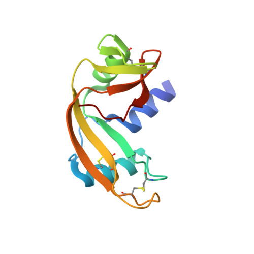
3ZBV, 3ZBW - PubMed Abstract:
Angiogenin (Ang) is a potent inducer of neovascularization. Point mutations in human Ang have been linked to cancer progression and two neurodegenerative diseases: amyotrophic lateral sclerosis and Parkinson's disease. Intensive structural and functional analyses of Ang have been paramount in assigning functions to this novel homologue of bovine pancreatic RNase A. However, inhibitor-binding studies with crystalline Ang (for designing potential anti-cancer drugs) have been hampered as a result of the inaccessibility of the active site. Experiments with the murine homologues of Ang have not only overcome the obvious practical limitations encountered when studying the role of a human protein in healthy individuals, but also the crystal structures of murine angiogenins (mAng and mAng-4) have revealed themselves to have greater potential for the visualization of small-molecule inhibitor binding at the active site. In the present study, we report the crystal structures of two more murine Ang paralogues, mAng-2 and mAng-3, at 1.6 and 1.8 Å resolution, respectively. These constitute the first crystal structures of an Ang with a zinc ion bound at the active site and provide some insight into the possible mode of inhibition of the ribonucleolytic activity of the enzyme by these divalent cations. Both structures show that the residues forming the putative P(1), B(1) and B(2) subsites occupy positions similar to their counterparts in human Ang and are likely to have conserved roles. However, a less obtrusive conformation of the C-terminal segment in mAng-3 and the presence of a sulfate ion in the B(1) subsite of mAng-2 suggest that these proteins have the potential to be used for inhibitor-binding studies. We also discuss the biological relevance of the structural similarities and differences between the different Ang homologues.
- Department of Biology and Biochemistry, University of Bath, Bath, UK.
Organizational Affiliation: