Electrophilic catalysis in triosephosphate isomerase: the role of histidine-95.
Komives, E.A., Chang, L.C., Lolis, E., Tilton, R.F., Petsko, G.A., Knowles, J.R.(1991) Biochemistry 30: 3011-3019
- PubMed: 2007138 Search on PubMed
- DOI: https://doi.org/10.1021/bi00226a005
- Primary Citation Related Structures:
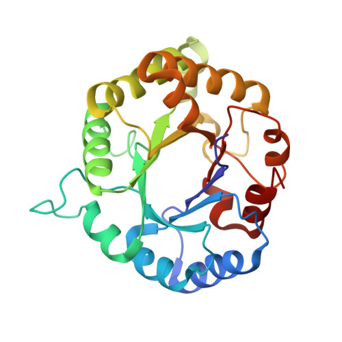
3YPI - PubMed Abstract:
Electrophilic catalysis by histidine-95 in triosephosphate isomerase has been probed by using Fourier transform infrared spectroscopy and X-ray crystallography. The carbonyl stretching frequency of dihydroxyacetone phosphate bound to the wild-type enzyme is known to be 19 cm-1 lower (at 1713 cm-1) than that of dihydroxyacetone phosphate free in solution (at 1732 cm-1), and this decrease in stretching frequency has been ascribed to an enzymic electrophile that polarizes the substrate carbonyl group toward the transition state for the enolization. Infrared spectra of substrate bound to two site-directed mutants of yeast triosephosphate isomerase in which histidine-95 has been changed to glutamine or to asparagine show unperturbed carbonyl stretching frequencies between 1732 and 1742 cm-1. The lack of carbonyl polarization when histidine-95 is removed suggests that histidine-95 is indeed the catalytic electrophile, at least for dihydroxyacetone phosphate. Kinetic studies of the glutamine mutant (H95Q) have shown that the enzyme follows a subtly different mechanism of proton transfers involving only a single acid-base catalytic group. These findings suggest an additional role for histidine-95 as a general acid-base catalyst in the wild-type enzyme. The X-ray crystal structure of the H95Q mutant with an intermediate analogue, phosphoglycolohydroxamate, bound at the active site has been solved to 2.8-A resolution, and this structure clearly implicates glutamate-165, the catalytic base in the wild-type isomerase, as the sole acid-base catalyst for the mutant enzyme.(ABSTRACT TRUNCATED AT 250 WORDS)
- Department of Chemistry, Harvard University, Cambridge, Massachusetts 02138.
Organizational Affiliation: