3X3B | pdb_00003x3b

Crystal structure of the light-driven sodium pump KR2 in acidic state


Experimental Data Snapshot

  • Method: X-RAY DIFFRACTION
  • Resolution: 2.30 Å
  • R-Value Free: 
    0.249 (Depositor), 0.252 (DCC) 
  • R-Value Work: 
    0.207 (Depositor), 0.214 (DCC) 
  • R-Value Observed: 
    0.211 (Depositor) 

Starting Model: experimental
View more details

wwPDB Validation 3D Report Full Report

Validation slider image for 3X3B

Ligand Structure Quality Assessment 


This is version 1.4 of the entry. See complete history

Literature

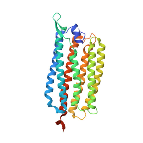
Structural basis for Na(+) transport mechanism by a light-driven Na(+) pump

Kato, H.E.Inoue, K.Abe-Yoshizumi, R.Kato, Y.Ono, H.Konno, M.Hososhima, S.Ishizuka, T.Hoque, M.R.Kunitomo, H.Ito, J.Yoshizawa, S.Yamashita, K.Takemoto, M.Nishizawa, T.Taniguchi, R.Kogure, K.Maturana, A.D.Iino, Y.Yawo, H.Ishitani, R.Kandori, H.Nureki, O.

(2015) Nature 521: 48-53

  • DOI: https://doi.org/10.1038/nature14322
  • Primary Citation Related Structures: 
    3X3B, 3X3C

  • PubMed Abstract: 

    Krokinobacter eikastus rhodopsin 2 (KR2) is the first light-driven Na(+) pump discovered, and is viewed as a potential next-generation optogenetics tool. Since the positively charged Schiff base proton, located within the ion-conducting pathway of all light-driven ion pumps, was thought to prohibit the transport of a non-proton cation, the discovery of KR2 raised the question of how it achieves Na(+) transport. Here we present crystal structures of KR2 under neutral and acidic conditions, which represent the resting and M-like intermediate states, respectively. Structural and spectroscopic analyses revealed the gating mechanism, whereby the flipping of Asp116 sequesters the Schiff base proton from the conducting pathway to facilitate Na(+) transport. Together with the structure-based engineering of the first light-driven K(+) pumps, electrophysiological assays in mammalian neurons and behavioural assays in a nematode, our studies reveal the molecular basis for light-driven non-proton cation pumps and thus provide a framework that may advance the development of next-generation optogenetics.


  • Organizational Affiliation
    • Department of Biological Sciences, Graduate School of Science, The University of Tokyo, 2-11-16 Yayoi, Bunkyo-ku, Tokyo 113-0032, Japan.

Macromolecule Content 

  • Total Structure Weight: 34.13 kDa 
  • Atom Count: 2,256 
  • Modeled Residue Count: 271 
  • Deposited Residue Count: 290 
  • Unique protein chains: 1

Macromolecules

Find similar proteins by:|  3D Structure
Entity ID: 1
MoleculeChains  Sequence LengthOrganismDetailsImage
Sodium pumping rhodopsin290Dokdonia eikastaMutation(s): 0 
Gene Names: NaR
Membrane Entity: Yes 
UniProt
Find proteins for N0DKS8 (Dokdonia eikasta)
Explore N0DKS8 
Go to UniProtKB:  N0DKS8
Entity Groups
Sequence Clusters30% Identity50% Identity70% Identity90% Identity95% Identity100% Identity
UniProt GroupN0DKS8
Sequence Annotations
Expand
Reference Sequence

Experimental Data & Validation

Experimental Data

  • Method: X-RAY DIFFRACTION
  • Resolution: 2.30 Å
  • R-Value Free:  0.249 (Depositor), 0.252 (DCC) 
  • R-Value Work:  0.207 (Depositor), 0.214 (DCC) 
  • R-Value Observed: 0.211 (Depositor) 
Space Group: I 2 2 2
Unit Cell:
Length ( Å )Angle ( ˚ )
a = 40.582α = 90
b = 81.544β = 90
c = 233.775γ = 90
Software Package:
Software NamePurpose
XDSdata scaling
MOLREPphasing
PHENIXrefinement
XDSdata reduction
SCALAdata scaling

Structure Validation

View Full Validation Report


Ligand Structure Quality Assessment 


Entry History 

Revision History  (Full details and data files)

  • Version 1.0: 2015-04-08
    Type: Initial release
  • Version 1.1: 2015-05-13
    Changes: Database references
  • Version 1.2: 2015-05-27
    Changes: Structure summary
  • Version 1.3: 2023-11-08
    Changes: Data collection, Database references, Derived calculations, Refinement description
  • Version 1.4: 2024-11-20
    Changes: Structure summary