The quaternary structure of the eukaryotic DNA replication proteins Sld7 and Sld3.
Itou, H., Shirakihara, Y., Araki, H.(2015) Acta Crystallogr D Biol Crystallogr 71: 1649-1656
- PubMed: 26249346 Search on PubMed
- DOI: https://doi.org/10.1107/S1399004715010457
- Primary Citation Related Structures:
3X37, 3X38 - PubMed Abstract:
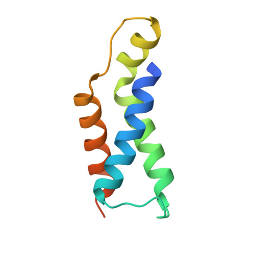
The initiation of eukaryotic chromosomal DNA replication requires the formation of an active replicative helicase at the replication origins of chromosomes. Yeast Sld3 and its metazoan counterpart treslin are the hub proteins mediating protein associations critical for formation of the helicase. The Sld7 protein interacts with Sld3, and the complex formed is thought to regulate the function of Sld3. Although Sld7 is a non-essential DNA replication protein that is found in only a limited range of yeasts, its depletion slowed the growth of cells and caused a delay in the S phase. Recently, the Mdm2-binding protein was found to bind to treslin in humans, and its depletion causes defects in cells similar to the depletion of Sld7 in yeast, suggesting their functional relatedness and importance during the initiation step of DNA replication. Here, the crystal structure of Sld7 in complex with Sld3 is presented. Sld7 comprises two structural domains. The N-terminal domain of Sld7 binds to Sld3, and the C-terminal domains connect two Sld7 molecules in an antiparallel manner. The quaternary structure of the Sld3-Sld7 complex shown from the crystal structures appears to be suitable to activate two helicase molecules loaded onto replication origins in a head-to-head manner.
- Structural Biology Center, National Institute of Genetics, Yata 1111, Mishima, Shizuoka 411-8540, Japan.
Organizational Affiliation: