3X32

Crystal structure of the oxidized form of the solubilized domain of porcine cytochrome b5 in form 1 crystal


Experimental Data Snapshot

  • Method: X-RAY DIFFRACTION
  • Resolution: 0.83 Å
  • R-Value Free: 0.127 
  • R-Value Work: 0.106 
  • R-Value Observed: 0.107 

wwPDB Validation   3D Report Full Report


Ligand Structure Quality Assessment 


This is version 1.3 of the entry. See complete history


Literature

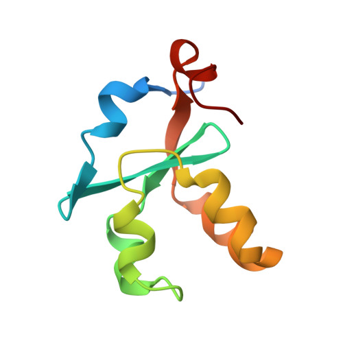
High-resolution crystal structures of the solubilized domain of porcine cytochrome b5.

Hirano, Y.Kimura, S.Tamada, T.

(2015) Acta Crystallogr D Biol Crystallogr 71: 1572-1581

  • DOI: https://doi.org/10.1107/S1399004715009438
  • Primary Citation of Related Structures:  
    3X32, 3X33, 3X34, 3X35

  • PubMed Abstract: 

    Mammalian microsomal cytochrome b5 has multiple electron-transfer partners that function in various electron-transfer reactions. Four crystal structures of the solubilized haem-binding domain of cytochrome b5 from porcine liver were determined at sub-angstrom resolution (0.76-0.95 Å) in two crystal forms for both the oxidized and reduced states. The high-resolution structures clearly displayed the electron density of H atoms in some amino-acid residues. Unrestrained refinement of bond lengths revealed that the protonation states of the haem propionate group may be involved in regulation of the haem redox properties. The haem Fe coordination geometry did not show significant differences between the oxidized and reduced structures. However, structural differences between the oxidized and reduced states were observed in the hydrogen-bond network around the axial ligand His68. The hydrogen-bond network could be involved in regulating the redox states of the haem group.


  • Organizational Affiliation

    Quantum Beam Science Center, Japan Atomic Energy Agency, 2-4 Shirakata, Tokai, Ibaraki 319-1195, Japan.


Macromolecules
Find similar proteins by:  (by identity cutoff)  |  3D Structure
Entity ID: 1
MoleculeChains Sequence LengthOrganismDetailsImage
Cytochrome b594Sus scrofaMutation(s): 0 
Gene Names: CYB5ACYB5
UniProt
Find proteins for P00172 (Sus scrofa)
Explore P00172 
Go to UniProtKB:  P00172
Entity Groups  
Sequence Clusters30% Identity50% Identity70% Identity90% Identity95% Identity100% Identity
UniProt GroupP00172
Sequence Annotations
Expand
  • Reference Sequence
Experimental Data & Validation

Experimental Data

  • Method: X-RAY DIFFRACTION
  • Resolution: 0.83 Å
  • R-Value Free: 0.127 
  • R-Value Work: 0.106 
  • R-Value Observed: 0.107 
  • Space Group: P 21 21 21
Unit Cell:
Length ( Å )Angle ( ˚ )
a = 32.339α = 90
b = 39.279β = 90
c = 60.95γ = 90
Software Package:
Software NamePurpose
SERGUIdata collection
PHASERphasing
SHELXL-97refinement
HKL-2000data reduction
HKL-2000data scaling

Structure Validation

View Full Validation Report


Ligand Structure Quality Assessment 


Entry History 

Deposition Data

Revision History  (Full details and data files)

  • Version 1.0: 2015-07-15
    Type: Initial release
  • Version 1.1: 2017-11-22
    Changes: Refinement description
  • Version 1.2: 2019-12-25
    Changes: Database references
  • Version 1.3: 2023-11-08
    Changes: Data collection, Database references, Derived calculations, Refinement description