3WVQ

Structure of ATP grasp protein


Experimental Data Snapshot

  • Method: X-RAY DIFFRACTION
  • Resolution: 1.96 Å
  • R-Value Free: 0.231 
  • R-Value Work: 0.194 
  • R-Value Observed: 0.196 

wwPDB Validation   3D Report Full Report


This is version 1.1 of the entry. See complete history


Literature

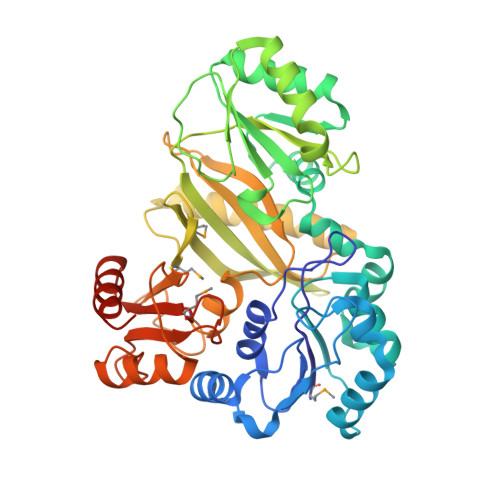
A peptide ligase and the ribosome cooperate to synthesize the peptide pheganomycin.

Noike, M.Matsui, T.Ooya, K.Sasaki, I.Ohtaki, S.Hamano, Y.Maruyama, C.Ishikawa, J.Satoh, Y.Ito, H.Morita, H.Dairi, T.

(2015) Nat Chem Biol 11: 71-76

  • DOI: https://doi.org/10.1038/nchembio.1697
  • Primary Citation of Related Structures:  
    3WVQ, 3WVR

  • PubMed Abstract: 

    Peptide antibiotics are typically biosynthesized by one of two distinct machineries in a ribosome-dependent or ribosome-independent manner. Pheganomycin (PGM (1)) and related analogs consist of the nonproteinogenic amino acid (S)-2-(3,5-dihydroxy-4-hydroxymethyl)phenyl-2-guanidinoacetic acid (2) and a proteinogenic core peptide, making their origin uncertain. We report the identification of the biosynthetic gene cluster from Streptomyces cirratus responsible for PGM production. Unexpectedly, the cluster contains a gene encoding multiple precursor peptides along with several genes plausibly encoding enzymes for the synthesis of amino acid 2. We identified PGM1, which has an ATP-grasp domain, as potentially capable of linking the precursor peptides with 2, and validate this hypothesis using deletion mutants and in vitro reconstitution. We document PGM1's substrate permissivity, which could be rationalized by a large binding pocket as confirmed via structural and mutagenesis experiments. This is to our knowledge the first example of cooperative peptide synthesis achieved by ribosomes and peptide ligases using a peptide nucleophile.


  • Organizational Affiliation

    Graduate School of Engineering, Hokkaido University, Hokkaido, Japan.


Macromolecules
Find similar proteins by:  (by identity cutoff)  |  3D Structure
Entity ID: 1
MoleculeChains Sequence LengthOrganismDetailsImage
PGM1
A, B, C, D
447Streptomyces cirratusMutation(s): 0 
UniProt
Find proteins for A0A0A6YVN3 (Streptomyces cirratus)
Explore A0A0A6YVN3 
Go to UniProtKB:  A0A0A6YVN3
Entity Groups  
Sequence Clusters30% Identity50% Identity70% Identity90% Identity95% Identity100% Identity
UniProt GroupA0A0A6YVN3
Sequence Annotations
Expand
  • Reference Sequence
Small Molecules
Ligands 2 Unique
IDChains Name / Formula / InChI Key2D Diagram3D Interactions
SO4
Query on SO4

Download Ideal Coordinates CCD File 
E [auth A]
F [auth A]
G [auth A]
H [auth B]
I [auth B]
E [auth A],
F [auth A],
G [auth A],
H [auth B],
I [auth B],
K [auth B],
L [auth B],
M [auth C],
N [auth C],
O [auth C],
P [auth C],
R [auth C],
S [auth C],
T [auth D],
U [auth D]
SULFATE ION
O4 S
QAOWNCQODCNURD-UHFFFAOYSA-L
GOL
Query on GOL

Download Ideal Coordinates CCD File 
J [auth B],
Q [auth C]
GLYCEROL
C3 H8 O3
PEDCQBHIVMGVHV-UHFFFAOYSA-N
Modified Residues  1 Unique
IDChains TypeFormula2D DiagramParent
MSE
Query on MSE
A, B, C, D
L-PEPTIDE LINKINGC5 H11 N O2 SeMET
Experimental Data & Validation

Experimental Data

  • Method: X-RAY DIFFRACTION
  • Resolution: 1.96 Å
  • R-Value Free: 0.231 
  • R-Value Work: 0.194 
  • R-Value Observed: 0.196 
  • Space Group: P 1
Unit Cell:
Length ( Å )Angle ( ˚ )
a = 77.66α = 73.74
b = 86.75β = 85.93
c = 94.26γ = 68.26
Software Package:
Software NamePurpose
XSCALEdata scaling
SOLVEphasing
PHENIXrefinement
PDB_EXTRACTdata extraction
XDSdata scaling
XDSdata reduction
PHASERphasing

Structure Validation

View Full Validation Report


Entry History 

Deposition Data

Revision History  (Full details and data files)

  • Version 1.0: 2014-11-19
    Type: Initial release
  • Version 1.1: 2014-12-31
    Changes: Database references