3WVC

Guanylylpyridinol (GP)-bound HcgF from Methanocaldococcus jannaschii


Experimental Data Snapshot

  • Method: X-RAY DIFFRACTION
  • Resolution: 2.00 Å
  • R-Value Free: 0.230 
  • R-Value Work: 0.163 
  • R-Value Observed: 0.166 

wwPDB Validation   3D Report Full Report


Ligand Structure Quality Assessment 


This is version 1.1 of the entry. See complete history


Literature

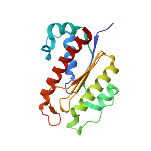
Protein-pyridinol thioester precursor for biosynthesis of the organometallic acyl-iron ligand in [Fe]-hydrogenase cofactor

Fujishiro, T.Kahnt, J.Ermler, U.Shima, S.

(2015) Nat Commun 6: 6895-6895

  • DOI: https://doi.org/10.1038/ncomms7895
  • Primary Citation of Related Structures:  
    3WV7, 3WV8, 3WV9, 3WVA, 3WVB, 3WVC

  • PubMed Abstract: 

    The iron-guanylylpyridinol (FeGP) cofactor of [Fe]-hydrogenase contains a prominent iron centre with an acyl-Fe bond and is the only acyl-organometallic iron compound found in nature. Here, we identify the functions of HcgE and HcgF, involved in the biosynthesis of the FeGP cofactor using structure-to-function strategy. Analysis of the HcgE and HcgF crystal structures with and without bound substrates suggest that HcgE catalyses the adenylylation of the carboxy group of guanylylpyridinol (GP) to afford AMP-GP, and subsequently HcgF catalyses the transesterification of AMP-GP to afford a Cys (HcgF)-S-GP thioester. Both enzymatic reactions are confirmed by in vitro assays. The structural data also offer plausible catalytic mechanisms. This strategy of thioester activation corresponds to that used for ubiquitin activation, a key event in the regulation of multiple cellular processes. It further implicates a nucleophilic attack onto the acyl carbon presumably via an electron-rich Fe(0)- or Fe(I)-carbonyl complex in the Fe-acyl formation.


  • Organizational Affiliation

    Max Planck Institute for Terrestrial Microbiology, Karl-von-Frisch-Strasse 10, 35043 Marburg, Germany.


Macromolecules
Find similar proteins by:  (by identity cutoff)  |  3D Structure
Entity ID: 1
MoleculeChains Sequence LengthOrganismDetailsImage
UPF0254 protein MJ1251
A, B
170Methanocaldococcus jannaschii DSM 2661Mutation(s): 0 
Gene Names: MJ1251
EC: 3.1.2
UniProt
Find proteins for Q58649 (Methanocaldococcus jannaschii (strain ATCC 43067 / DSM 2661 / JAL-1 / JCM 10045 / NBRC 100440))
Explore Q58649 
Go to UniProtKB:  Q58649
Entity Groups  
Sequence Clusters30% Identity50% Identity70% Identity90% Identity95% Identity100% Identity
UniProt GroupQ58649
Sequence Annotations
Expand
  • Reference Sequence
Small Molecules
Ligands 3 Unique
IDChains Name / Formula / InChI Key2D Diagram3D Interactions
ZGP
Query on ZGP

Download Ideal Coordinates CCD File 
E [auth A]5'-O-[(R)-hydroxy{[2-hydroxy-3,5-dimethyl-6-(2-oxo-2-sulfanylethyl)pyridin-4-yl]oxy}phosphoryl]guanosine
C19 H23 N6 O10 P S
JZNXQNYXQOKUIF-ACDAHVMNSA-N
FEG
Query on FEG

Download Ideal Coordinates CCD File 
F [auth B]5'-O-[(S)-{[2-(carboxymethyl)-6-hydroxy-3,5-dimethylpyridin-4-yl]oxy}(hydroxy)phosphoryl]guanosine
C19 H23 N6 O11 P
GTHOTKPHMGEHFD-ACDAHVMNSA-N
SO4
Query on SO4

Download Ideal Coordinates CCD File 
C [auth A]
D [auth A]
G [auth B]
H [auth B]
I [auth B]
C [auth A],
D [auth A],
G [auth B],
H [auth B],
I [auth B],
J [auth B]
SULFATE ION
O4 S
QAOWNCQODCNURD-UHFFFAOYSA-L
Experimental Data & Validation

Experimental Data

  • Method: X-RAY DIFFRACTION
  • Resolution: 2.00 Å
  • R-Value Free: 0.230 
  • R-Value Work: 0.163 
  • R-Value Observed: 0.166 
  • Space Group: P 1 21 1
Unit Cell:
Length ( Å )Angle ( ˚ )
a = 39.57α = 90
b = 97.53β = 111.17
c = 47.12γ = 90
Software Package:
Software NamePurpose
XSCALEdata scaling
REFMACrefinement
PDB_EXTRACTdata extraction
XDSdata scaling
XDSdata reduction
MOLREPphasing

Structure Validation

View Full Validation Report


Ligand Structure Quality Assessment 


Entry History 

Deposition Data

Revision History  (Full details and data files)

  • Version 1.0: 2015-04-29
    Type: Initial release
  • Version 1.1: 2023-11-08
    Changes: Data collection, Database references, Derived calculations, Refinement description