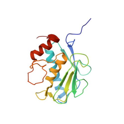
3WV2

Crystal structure of the catalytic domain of MMP-13 complexed with N-(3-methoxybenzyl)-4-oxo-3,4-dihydroquinazoline-2-carboxamide


Experimental Data Snapshot

  • Method: X-RAY DIFFRACTION
  • Resolution: 2.30 Å
  • R-Value Free: 0.241 
  • R-Value Work: 0.171 
  • R-Value Observed: 0.174 

wwPDB Validation   3D Report Full Report


Ligand Structure Quality Assessment 


This is version 1.2 of the entry. See complete history


Literature

Thieno[2,3-d]pyrimidine-2-carboxamides bearing a carboxybenzene group at 5-position: highly potent, selective, and orally available MMP-13 inhibitors interacting with the S1′′ binding site.

Nara, H.Sato, K.Naito, T.Mototani, H.Oki, H.Yamamoto, Y.Kuno, H.Santou, T.Kanzaki, N.Terauchi, J.Uchikawa, O.Kori, M.

(2014) Bioorg Med Chem 22: 5487-5505

  • DOI: https://doi.org/10.1016/j.bmc.2014.07.025
  • Primary Citation of Related Structures:  
    3WV2, 3WV3

  • PubMed Abstract: 

    On the basis of X-ray co-crystal structures of matrix metalloproteinase-13 (MMP-13) in complex with its inhibitors, our structure-based drug design (SBDD) strategy was directed to achieving high affinity through optimal protein-ligand interaction with the unique S1″ hydrophobic specificity pocket. This report details the optimization of lead compound 44 to highly potent and selective MMP-13 inhibitors based on fused pyrimidine scaffolds represented by the thienopyrimidin-4-one 26c. Furthermore, we have examined the release of collagen fragments from bovine nasal cartilage in response to a combination of IL-1 and oncostatin M.


  • Organizational Affiliation

    Pharmaceutical Research Division, Takeda Pharmaceutical Company Ltd, 26-1, Muraokahigashi 2-Chome, Fujisawa, Kanagawa 251-8555, Japan. Electronic address: hiroshi.nara@takeda.com.


Macromolecules
Find similar proteins by:  (by identity cutoff)  |  3D Structure
Entity ID: 1
MoleculeChains Sequence LengthOrganismDetailsImage
Collagenase 3
A, B
171Homo sapiensMutation(s): 0 
Gene Names: MMP13
EC: 3.4.24
UniProt & NIH Common Fund Data Resources
Find proteins for P45452 (Homo sapiens)
Explore P45452 
Go to UniProtKB:  P45452
PHAROS:  P45452
GTEx:  ENSG00000137745 
Entity Groups  
Sequence Clusters30% Identity50% Identity70% Identity90% Identity95% Identity100% Identity
UniProt GroupP45452
Sequence Annotations
Expand
  • Reference Sequence
Small Molecules
Ligands 4 Unique
IDChains Name / Formula / InChI Key2D Diagram3D Interactions
WGG
Query on WGG

Download Ideal Coordinates CCD File 
H [auth A],
N [auth B]
N-(3-methoxybenzyl)-4-oxo-3,4-dihydroquinazoline-2-carboxamide
C17 H15 N3 O3
AARSEFXVCWMMKV-UHFFFAOYSA-N
ZN
Query on ZN

Download Ideal Coordinates CCD File 
C [auth A],
D [auth A],
I [auth B],
J [auth B]
ZINC ION
Zn
PTFCDOFLOPIGGS-UHFFFAOYSA-N
CA
Query on CA

Download Ideal Coordinates CCD File 
E [auth A],
F [auth A],
K [auth B],
L [auth B]
CALCIUM ION
Ca
BHPQYMZQTOCNFJ-UHFFFAOYSA-N
NA
Query on NA

Download Ideal Coordinates CCD File 
G [auth A],
M [auth B]
SODIUM ION
Na
FKNQFGJONOIPTF-UHFFFAOYSA-N
Binding Affinity Annotations 
IDSourceBinding Affinity
WGG BindingDB:  3WV2 IC50: min: 5.6, max: 12 (nM) from 2 assay(s)
Experimental Data & Validation

Experimental Data

  • Method: X-RAY DIFFRACTION
  • Resolution: 2.30 Å
  • R-Value Free: 0.241 
  • R-Value Work: 0.171 
  • R-Value Observed: 0.174 
  • Space Group: C 1 2 1
Unit Cell:
Length ( Å )Angle ( ˚ )
a = 143.53α = 90
b = 35.719β = 135.29
c = 94.762γ = 90
Software Package:
Software NamePurpose
d*TREKdata reduction
REFMACrefinement
PDB_EXTRACTdata extraction
CrystalCleardata collection
CrystalCleardata reduction
CrystalCleardata scaling
MOLREPphasing

Structure Validation

View Full Validation Report


Ligand Structure Quality Assessment 


Entry History 

Deposition Data

Revision History  (Full details and data files)

  • Version 1.0: 2014-09-24
    Type: Initial release
  • Version 1.1: 2022-08-24
    Changes: Database references, Derived calculations
  • Version 1.2: 2024-05-29
    Changes: Data collection