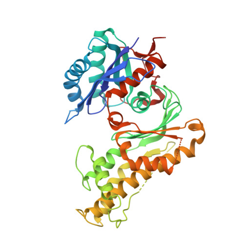
3WUH

Qri7 and AMP complex


Experimental Data Snapshot

  • Method: X-RAY DIFFRACTION
  • Resolution: 2.94 Å
  • R-Value Free: 0.273 
  • R-Value Work: 0.223 
  • R-Value Observed: 0.226 

Starting Model: experimental
View more details

wwPDB Validation   3D Report Full Report


Ligand Structure Quality Assessment 


This is version 1.1 of the entry. See complete history


Literature

Structure of Saccharomyces cerevisiae mitochondrial Qri7 in complex with AMP

Tominaga, T.Kobayashi, K.Ishii, R.Ishitani, R.Nureki, O.

(2014) Acta Crystallogr F Struct Biol Commun 70: 1009-1014

  • DOI: https://doi.org/10.1107/S2053230X14014046
  • Primary Citation of Related Structures:  
    3WUH

  • PubMed Abstract: 

    N(6)-Threonylcarbamoyladenosine (t(6)A) is a modified tRNA base required for accuracy in translation. Qri7 is localized in yeast mitochondria and is involved in t(6)A biosynthesis. In t(6)A biosynthesis, threonylcarbamoyl-adenylate (TCA) is synthesized from threonine, bicarbonate and ATP, and the threonyl-carbamoyl group is transferred to adenine 37 of tRNA by Qri7. Qri7 alone is sufficient to catalyze the second step of the reaction, whereas the Qri7 homologues YgjD (in bacteria) and Kae1 (in archaea and eukaryotes) function as parts of multi-protein complexes. In this study, the crystal structure of Qri7 complexed with AMP (a part of TCA) has been determined at 2.94 Å resolution in a new crystal form. The manner of AMP recognition is similar, with some minor variations, among the Qri7/Kae1/YgjD family proteins. The previously reported dimer formation was also observed in this new crystal form. Furthermore, a comparison with the structure of TobZ, which catalyzes a similar reaction to t(6)A biosynthesis, revealed the presence of a flexible loop that may be involved in tRNA binding.


  • Organizational Affiliation

    Department of Biological Sciences, Graduate School of Science, The University of Tokyo, 2-11-16 Yayoi, Bunkyo-ku, Tokyo 113-0032, Japan.


Macromolecules
Find similar proteins by:  (by identity cutoff)  |  3D Structure
Entity ID: 1
MoleculeChains Sequence LengthOrganismDetailsImage
tRNA N6-adenosine threonylcarbamoyltransferase, mitochondrial
A, B
383Saccharomyces cerevisiae S288CMutation(s): 0 
Gene Names: QRI7YDL104CD2366
EC: 2.3.1
UniProt
Find proteins for P43122 (Saccharomyces cerevisiae (strain ATCC 204508 / S288c))
Explore P43122 
Go to UniProtKB:  P43122
Entity Groups  
Sequence Clusters30% Identity50% Identity70% Identity90% Identity95% Identity100% Identity
UniProt GroupP43122
Sequence Annotations
Expand
  • Reference Sequence
Experimental Data & Validation

Experimental Data

  • Method: X-RAY DIFFRACTION
  • Resolution: 2.94 Å
  • R-Value Free: 0.273 
  • R-Value Work: 0.223 
  • R-Value Observed: 0.226 
  • Space Group: P 43 3 2
Unit Cell:
Length ( Å )Angle ( ˚ )
a = 180.315α = 90
b = 180.315β = 90
c = 180.315γ = 90
Software Package:
Software NamePurpose
HKL-2000data collection
PHASERphasing
PHENIXrefinement
HKL-2000data reduction
HKL-2000data scaling

Structure Validation

View Full Validation Report


Ligand Structure Quality Assessment 


Entry History 

Deposition Data

Revision History  (Full details and data files)

  • Version 1.0: 2014-09-17
    Type: Initial release
  • Version 1.1: 2023-11-08
    Changes: Data collection, Database references, Derived calculations, Refinement description