A redox switch shapes the Lon protease exit pore to facultatively regulate proteolysis.
Nishii, W., Kukimoto-Niino, M., Terada, T., Shirouzu, M., Muramatsu, T., Kojima, M., Kihara, H., Yokoyama, S.(2015) Nat Chem Biol 11: 46-51
- PubMed: 25383757 Search on PubMed
- DOI: https://doi.org/10.1038/nchembio.1688
- Primary Citation Related Structures:
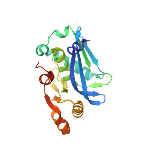
3WU3, 3WU4, 3WU5, 3WU6 - PubMed Abstract:
The Lon AAA+ protease degrades damaged or misfolded proteins in its intramolecular chamber. Its activity must be precisely controlled, but the mechanism by which Lon is regulated in response to different environments is not known. Facultative anaerobes in the Enterobacteriaceae family, mostly symbionts and pathogens, encounter both anaerobic and aerobic environments inside and outside the host's body, respectively. The bacteria characteristically have two cysteine residues on the Lon protease (P) domain surface that unusually form a disulfide bond. Here we show that the cysteine residues act as a redox switch of Lon. Upon disulfide bond reduction, the exit pore of the P-domain ring narrows by ∼30%, thus interrupting product passage and decreasing activity by 80%; disulfide bonding by oxidation restores the pore size and activity. The redox switch (E°' = -227 mV) is appropriately tuned to respond to variation between anaerobic and aerobic conditions, thus optimizing the cellular proteolysis level for each environment.
- 1] RIKEN Systems and Structural Biology Center, Yokohama, Japan. [2] RIKEN Structural Biology Laboratory, Yokohama, Japan.
Organizational Affiliation: