


Structural basis on the efficient CO2 reduction of acidophilic formate dehydrogenase
Ha, J.M., Jeon, S.T., Yoon, H.J., Lee, H.H.To be published.
Experimental Data Snapshot
Starting Model: experimental
View more details
Entity ID: 1 | |||||
|---|---|---|---|---|---|
| Molecule | Chains | Sequence Length | Organism | Details | Image |
| Formate dehydrogenase | 406 | Thiobacillus sp. KNK65MA | Mutation(s): 0 Gene Names: fdh65MA EC: 1.2.1.2 (PDB Primary Data), 1.17.1.9 (UniProt) | 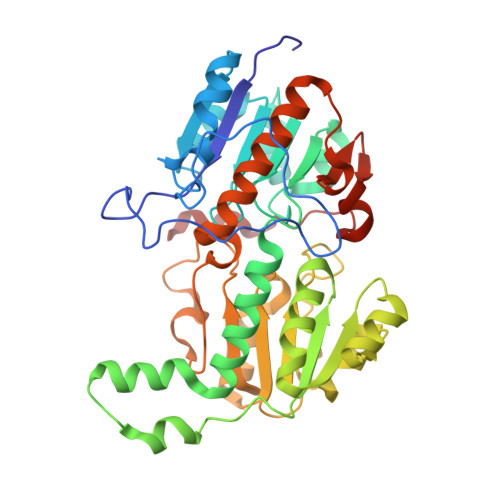 | |
UniProt | |||||
Entity Groups | |||||
| Sequence Clusters | 30% Identity50% Identity70% Identity90% Identity95% Identity100% Identity | ||||
| UniProt Group | Q76EB7 | ||||
Sequence AnnotationsExpand | |||||
Reference Sequence | |||||
| Ligands 1 Unique | |||||
|---|---|---|---|---|---|
| ID | Chains | Name / Formula / InChI Key | 2D Diagram | 3D Interactions | |
| NAD Download:Ideal Coordinates CCD File | E [auth A], F [auth B], G [auth C] | NICOTINAMIDE-ADENINE-DINUCLEOTIDE C21 H27 N7 O14 P2 BAWFJGJZGIEFAR-NNYOXOHSSA-N |  | ||
| Length ( Å ) | Angle ( ˚ ) |
|---|---|
| a = 55.942 | α = 90 |
| b = 72.729 | β = 90 |
| c = 386.767 | γ = 90 |
| Software Name | Purpose |
|---|---|
| HKL-2000 | data collection |
| PHENIX | model building |
| PHENIX | refinement |
| DENZO | data reduction |
| HKL-2000 | data scaling |
| PHENIX | phasing |


