3WKE | pdb_00003wke

Crystal structure of soluble epoxide hydrolase in complex with t-AUCB


Experimental Data Snapshot

  • Method: X-RAY DIFFRACTION
  • Resolution: 2.75 Å
  • R-Value Free: 
    0.254 (Depositor), 0.254 (DCC) 
  • R-Value Work: 
    0.174 (Depositor), 0.174 (DCC) 
  • R-Value Observed: 
    0.178 (Depositor) 

wwPDB Validation 3D Report Full Report

Validation slider image for 3WKE

Ligand Structure Quality Assessment 


This is version 1.2 of the entry. See complete history

Literature

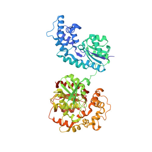
Structural insights into binding of inhibitors to soluble epoxide hydrolase gained by fragment screening and X-ray crystallography.

Amano, Y.Yamaguchi, T.Tanabe, E.

(2014) Bioorg Med Chem 22: 2427-2434

  • DOI: https://doi.org/10.1016/j.bmc.2014.03.001
  • Primary Citation Related Structures: 
    3WK4, 3WK5, 3WK6, 3WK7, 3WK8, 3WK9, 3WKA, 3WKB, 3WKC, 3WKD, 3WKE

  • PubMed Abstract: 

    Soluble epoxide hydrolase (sEH) is a component of the arachidonic acid cascade and is a candidate target for therapies for hypertension or inflammation. Although many sEH inhibitors are available, their scaffolds are not structurally diverse, and knowledge of their specific interactions with sEH is limited. To obtain detailed structural information about protein-ligand interactions, we conducted fragment screening of sEH, analyzed the fragments using high-throughput X-ray crystallography, and determined 126 fragment-bound structures at high resolution. Aminothiazole and benzimidazole derivatives were identified as novel scaffolds that bind to the catalytic triad of sEH with good ligand efficiency. We further identified fragment hits that bound to subpockets of sEH called the short and long branches. The water molecule conserved in the structure plays an important role in binding to the long branch, whereas Asp496 and the main chain of Phe497 form hydrogen bonds with fragment hits in the short branch. Fragment hits and their crystal structures provide structural insights into ligand binding to sEH that will facilitate the discovery of novel and potent inhibitors of sEH.


  • Organizational Affiliation
    • Drug Discovery Research, Astellas Pharma Inc., 21, Miyukigaoka, Tsukuba, Ibaraki, Japan. Electronic address: yasushi-amano@astellas.com.

Macromolecule Content 

  • Total Structure Weight: 64.05 kDa 
  • Atom Count: 4,440 
  • Modeled Residue Count: 546 
  • Deposited Residue Count: 561 
  • Unique protein chains: 1

Macromolecules

Find similar proteins by:|  3D Structure
Entity ID: 1
MoleculeChains  Sequence LengthOrganismDetailsImage
Bifunctional epoxide hydrolase 2561Homo sapiensMutation(s): 0 
Gene Names: EPHX2
EC: 3.3.2.10 (PDB Primary Data), 3.1.3.76 (PDB Primary Data)
UniProt & NIH Common Fund Data Resources
Find proteins for P34913 (Homo sapiens)
Explore P34913 
Go to UniProtKB:  P34913
PHAROS:  P34913
GTEx:  ENSG00000120915 
Entity Groups
Sequence Clusters30% Identity50% Identity70% Identity90% Identity95% Identity100% Identity
UniProt GroupP34913
Sequence Annotations
Expand
Reference Sequence

Small Molecules

Ligands 3 Unique
IDChains Name / Formula / InChI Key2D Diagram3D Interactions
AUB

Query on AUB


Download:Ideal Coordinates CCD File
D [auth A]4-[(trans-4-{[(3s,5s,7s)-tricyclo[3.3.1.1~3,7~]dec-1-ylcarbamoyl]amino}cyclohexyl)oxy]benzoic acid
C24 H32 N2 O4
KNBWKJBQDAQARU-GNCKNZPZSA-N
PO4

Query on PO4


Download:Ideal Coordinates CCD File
C [auth A]PHOSPHATE ION
O4 P
NBIIXXVUZAFLBC-UHFFFAOYSA-K
MG

Query on MG


Download:Ideal Coordinates CCD File
B [auth A]MAGNESIUM ION
Mg
JLVVSXFLKOJNIY-UHFFFAOYSA-N

Experimental Data & Validation

Experimental Data

  • Method: X-RAY DIFFRACTION
  • Resolution: 2.75 Å
  • R-Value Free:  0.254 (Depositor), 0.254 (DCC) 
  • R-Value Work:  0.174 (Depositor), 0.174 (DCC) 
  • R-Value Observed: 0.178 (Depositor) 
Space Group: P 65 2 2
Unit Cell:
Length ( Å )Angle ( ˚ )
a = 91.948α = 90
b = 91.948β = 90
c = 243.942γ = 120
Software Package:
Software NamePurpose
ADSCdata collection
AMoREphasing
REFMACrefinement
HKL-2000data reduction
HKL-2000data scaling

Structure Validation

View Full Validation Report


Ligand Structure Quality Assessment 


Entry History 

Deposition Data

Revision History  (Full details and data files)

  • Version 1.0: 2014-04-16
    Type: Initial release
  • Version 1.1: 2022-08-24
    Changes: Database references, Derived calculations
  • Version 1.2: 2024-05-29
    Changes: Data collection