3WI2 | pdb_00003wi2

Crystal structure of PDE10A in complex with inhibitor


Experimental Data Snapshot

  • Method: X-RAY DIFFRACTION
  • Resolution: 2.26 Å
  • R-Value Free: 
    0.329 (Depositor), 0.328 (DCC) 
  • R-Value Work: 
    0.253 (Depositor), 0.252 (DCC) 
  • R-Value Observed: 
    0.257 (Depositor) 

wwPDB Validation 3D Report Full Report

Validation slider image for 3WI2

Ligand Structure Quality Assessment 


This is version 1.2 of the entry. See complete history

Literature

Design and synthesis of novel benzimidazole derivatives as phosphodiesterase 10A inhibitors with reduced CYP1A2 inhibition.

Hamaguchi, W.Masuda, N.Isomura, M.Miyamoto, S.Kikuchi, S.Amano, Y.Honbou, K.Mihara, T.Watanabe, T.

(2013) Bioorg Med Chem 21: 7612-7623

  • DOI: https://doi.org/10.1016/j.bmc.2013.10.035
  • Primary Citation Related Structures: 
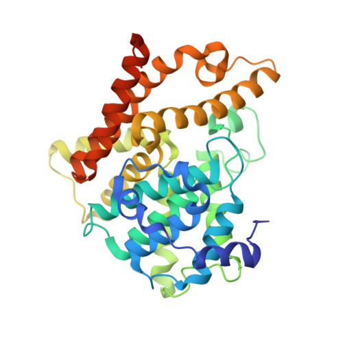
    3WI2

  • PubMed Abstract: 

    A novel class of phosphodiesterase 10A (PDE10A) inhibitors with reduced CYP1A2 inhibition were designed and synthesized starting from 2-{[(1-phenyl-1H-benzimidazol-6-yl)oxy]methyl}quinoline (1). Introduction of an isopropyl group at the 2-position and a methoxy group at the 5-position of the benzimidazole ring of lead compound 1 resulted in the identification of 2-{[(2-isopropyl-5-methoxy-1-phenyl-1H-benzimidazol-6-yl)oxy]methyl}quinoline (25b), which exhibited potent PDE10A inhibitory activity with reduced CYP1A2 inhibitory activity compared to compound 1.


  • Organizational Affiliation
    • Drug Discovery Research, Astellas Pharma Inc., 21, Miyukigaoka, Tsukuba-shi, Ibaraki 305-8585, Japan. Electronic address: wataru.hamaguchi@astellas.com.

Macromolecule Content 

  • Total Structure Weight: 80.18 kDa 
  • Atom Count: 5,335 
  • Modeled Residue Count: 648 
  • Deposited Residue Count: 690 
  • Unique protein chains: 1

Macromolecules

Find similar proteins by:|  3D Structure
Entity ID: 1
MoleculeChains  Sequence LengthOrganismDetailsImage
cAMP and cAMP-inhibited cGMP 3',5'-cyclic phosphodiesterase 10A
A, B
345Homo sapiensMutation(s): 0 
Gene Names: PDE10A
EC: 3.1.4.17 (PDB Primary Data), 3.1.4.35 (PDB Primary Data)
UniProt & NIH Common Fund Data Resources
Find proteins for Q9Y233 (Homo sapiens)
Explore Q9Y233 
Go to UniProtKB:  Q9Y233
PHAROS:  Q9Y233
GTEx:  ENSG00000112541 
Entity Groups
Sequence Clusters30% Identity50% Identity70% Identity90% Identity95% Identity100% Identity
UniProt GroupQ9Y233
Sequence Annotations
Expand
Reference Sequence

Experimental Data & Validation

Experimental Data

  • Method: X-RAY DIFFRACTION
  • Resolution: 2.26 Å
  • R-Value Free:  0.329 (Depositor), 0.328 (DCC) 
  • R-Value Work:  0.253 (Depositor), 0.252 (DCC) 
  • R-Value Observed: 0.257 (Depositor) 
Space Group: P 21 21 21
Unit Cell:
Length ( Å )Angle ( ˚ )
a = 50.115α = 90
b = 81.629β = 90
c = 158.084γ = 90
Software Package:
Software NamePurpose
CrystalCleardata collection
AMoREphasing
REFMACrefinement
CrystalCleardata reduction
CrystalCleardata scaling

Structure Validation

View Full Validation Report


Ligand Structure Quality Assessment 


Entry History 

Deposition Data

  • Released Date: 2013-12-11 
  • Deposition Author(s): Amano, Y.

Revision History  (Full details and data files)

  • Version 1.0: 2013-12-11
    Type: Initial release
  • Version 1.1: 2022-08-24
    Changes: Database references, Derived calculations
  • Version 1.2: 2024-05-29
    Changes: Data collection