3WEJ | pdb_00003wej

Crystal structure of the human squalene synthase F288A mutant in complex with presqualene pyrophosphate


Experimental Data Snapshot

  • Method: X-RAY DIFFRACTION
  • Resolution: 2.00 Å
  • R-Value Free: 
    0.276 (Depositor), 0.275 (DCC) 
  • R-Value Work: 
    0.208 (Depositor), 0.206 (DCC) 
  • R-Value Observed: 
    0.211 (Depositor) 

Starting Model: experimental
View more details

wwPDB Validation 3D Report Full Report

Validation slider image for 3WEJ

Ligand Structure Quality Assessment 


This is version 1.2 of the entry. See complete history

Literature

Structural insights into the catalytic mechanism of human squalene synthase.

Liu, C.I.Jeng, W.Y.Chang, W.J.Shih, M.F.Ko, T.P.Wang, A.H.J.

(2014) Acta Crystallogr D Biol Crystallogr 70: 231-241

  • DOI: https://doi.org/10.1107/S1399004713026230
  • Primary Citation Related Structures: 
    3WEF, 3WEG, 3WEH, 3WEI, 3WEJ, 3WEK

  • PubMed Abstract: 

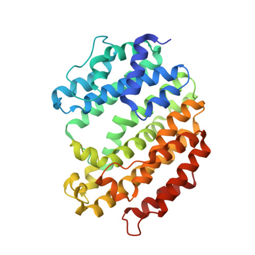
    Squalene synthase (SQS) is a divalent metal-ion-dependent enzyme that catalyzes the two-step reductive `head-to-head' condensation of two molecules of farnesyl pyrophosphate to form squalene using presqualene diphosphate (PSPP) as an intermediate. In this paper, the structures of human SQS and its mutants in complex with several substrate analogues and intermediates coordinated with Mg2+ or Mn2+ are presented, which stepwise delineate the biosynthetic pathway. Extensive study of the SQS active site has identified several critical residues that are involved in binding reduced nicotinamide dinucleotide phosphate (NADPH). Based on mutagenesis data and a locally closed (JK loop-in) structure observed in the hSQS-(F288L)-PSPP complex, an NADPH-binding model is proposed for SQS. The results identified four major steps (substrate binding, condensation, intermediate formation and translocation) of the ordered sequential mechanisms involved in the `1'-1' isoprenoid biosynthetic pathway. These new findings clarify previous hypotheses based on site-directed mutagenesis and biochemical analysis.


  • Organizational Affiliation
    • Institute of Biological Chemistry, Academia Sinica, Taipei 11529, Taiwan.

Macromolecule Content 

  • Total Structure Weight: 40.01 kDa 
  • Atom Count: 2,989 
  • Modeled Residue Count: 335 
  • Deposited Residue Count: 343 
  • Unique protein chains: 1

Macromolecules

Find similar proteins by:|  3D Structure
Entity ID: 1
MoleculeChains  Sequence LengthOrganismDetailsImage
Squalene synthase343Homo sapiensMutation(s): 1 
Gene Names: FDFT1
EC: 2.5.1.21
UniProt & NIH Common Fund Data Resources
Find proteins for P37268 (Homo sapiens)
Explore P37268 
Go to UniProtKB:  P37268
PHAROS:  P37268
GTEx:  ENSG00000079459 
Entity Groups
Sequence Clusters30% Identity50% Identity70% Identity90% Identity95% Identity100% Identity
UniProt GroupP37268
Sequence Annotations
Expand
Reference Sequence

Small Molecules

Ligands 2 Unique
IDChains Name / Formula / InChI Key2D Diagram3D Interactions
PS7

Query on PS7


Download:Ideal Coordinates CCD File
D [auth A]{(1R,2R,3R)-2-[(3E)-4,8-dimethylnona-3,7-dien-1-yl]-2-methyl-3-[(1E,5E)-2,6,10-trimethylundeca-1,5,9-trien-1-yl]cyclopropyl}methyl trihydrogen diphosphate
C30 H52 O7 P2
ATZKAUGGNMSCCY-VVFNRDJMSA-N
MG

Query on MG


Download:Ideal Coordinates CCD File
B [auth A],
C [auth A]
MAGNESIUM ION
Mg
JLVVSXFLKOJNIY-UHFFFAOYSA-N

Experimental Data & Validation

Experimental Data

  • Method: X-RAY DIFFRACTION
  • Resolution: 2.00 Å
  • R-Value Free:  0.276 (Depositor), 0.275 (DCC) 
  • R-Value Work:  0.208 (Depositor), 0.206 (DCC) 
  • R-Value Observed: 0.211 (Depositor) 
Space Group: P 21 21 21
Unit Cell:
Length ( Å )Angle ( ˚ )
a = 35.714α = 90
b = 94.503β = 90
c = 105.314γ = 90
Software Package:
Software NamePurpose
HKL-2000data collection
MOLREPphasing
REFMACrefinement
HKL-2000data reduction
HKL-2000data scaling

Structure Validation

View Full Validation Report


Ligand Structure Quality Assessment 


Entry History 

Deposition Data

Revision History  (Full details and data files)

  • Version 1.0: 2014-02-12
    Type: Initial release
  • Version 1.1: 2019-12-25
    Changes: Data collection, Database references
  • Version 1.2: 2023-11-08
    Changes: Data collection, Database references, Derived calculations, Refinement description