3WC2

Crystal structure of C. albicans tRNA(His) guanylyltransferase (Thg1) with a tRNA(Phe)(GUG)


Experimental Data Snapshot

  • Method: X-RAY DIFFRACTION
  • Resolution: 3.64 Å
  • R-Value Free: 0.287 
  • R-Value Work: 0.253 
  • R-Value Observed: 0.255 

wwPDB Validation   3D Report Full Report


This is version 1.4 of the entry. See complete history


Literature

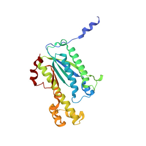
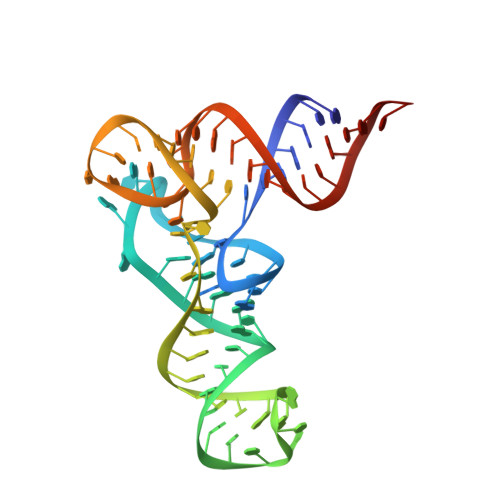
Structural basis of reverse nucleotide polymerization

Nakamura, A.Nemoto, T.Heinemann, I.U.Yamashita, K.Sonoda, T.Komoda, K.Tanaka, I.Soll, D.Yao, M.

(2013) Proc Natl Acad Sci U S A 110: 20970-20975

  • DOI: https://doi.org/10.1073/pnas.1321312111
  • Primary Citation of Related Structures:  
    3WBZ, 3WC0, 3WC1, 3WC2

  • PubMed Abstract: 

    Nucleotide polymerization proceeds in the forward (5'-3') direction. This tenet of the central dogma of molecular biology is found in diverse processes including transcription, reverse transcription, DNA replication, and even in lagging strand synthesis where reverse polymerization (3'-5') would present a "simpler" solution. Interestingly, reverse (3'-5') nucleotide addition is catalyzed by the tRNA maturation enzyme tRNA(His) guanylyltransferase, a structural homolog of canonical forward polymerases. We present a Candida albicans tRNA(His) guanylyltransferase-tRNA(His) complex structure that reveals the structural basis of reverse polymerization. The directionality of nucleotide polymerization is determined by the orientation of approach of the nucleotide substrate. The tRNA substrate enters the enzyme's active site from the opposite direction (180° flip) compared with similar nucleotide substrates of canonical 5'-3' polymerases, and the finger domains are on opposing sides of the core palm domain. Structural, biochemical, and phylogenetic data indicate that reverse polymerization appeared early in evolution and resembles a mirror image of the forward process.


  • Organizational Affiliation

    Faculty of Advanced Life Science, Hokkaido University, Sapporo 060-0810, Japan.


Macromolecules

Find similar proteins by:  (by identity cutoff)  |  3D Structure
Entity ID: 1
MoleculeChains Sequence LengthOrganismDetailsImage
Likely histidyl tRNA-specific guanylyltransferase
A, B, C, D
271Candida albicans SC5314Mutation(s): 0 
Gene Names: CaO19.7063CAWG_05412orf19.7063THG1
Entity Groups  
Sequence Clusters30% Identity50% Identity70% Identity90% Identity95% Identity100% Identity
Sequence Annotations
Expand
  • Reference Sequence
Find similar nucleic acids by:  (by identity cutoff)  |  3D Structure
Entity ID: 2
MoleculeChains LengthOrganismImage
76mer-tRNAE [auth P],
F [auth Q]
76N/A
Sequence Annotations
Expand
  • Reference Sequence
Experimental Data & Validation

Experimental Data

  • Method: X-RAY DIFFRACTION
  • Resolution: 3.64 Å
  • R-Value Free: 0.287 
  • R-Value Work: 0.253 
  • R-Value Observed: 0.255 
  • Space Group: P 31 2 1
Unit Cell:
Length ( Å )Angle ( ˚ )
a = 96.74α = 90
b = 96.74β = 90
c = 299.36γ = 120
Software Package:
Software NamePurpose
XSCALEdata scaling
PHENIXrefinement
PDB_EXTRACTdata extraction
SERGUIdata collection
XDSdata reduction
XDSdata scaling
PHASERphasing

Structure Validation

View Full Validation Report


Entry History 

Deposition Data

Revision History  (Full details and data files)

  • Version 1.0: 2013-12-18
    Type: Initial release
  • Version 1.1: 2014-01-15
    Changes: Database references
  • Version 1.2: 2017-11-22
    Changes: Refinement description
  • Version 1.3: 2019-12-18
    Changes: Data collection, Database references
  • Version 1.4: 2023-11-08
    Changes: Data collection, Database references, Refinement description