3WAE

X-ray structure of Fe(III)-bicarbonates-ttfbpa, a ferric ion-binding protein from thermus thermophilus HB8


Experimental Data Snapshot

  • Method: X-RAY DIFFRACTION
  • Resolution: 1.70 Å
  • R-Value Free: 0.243 
  • R-Value Work: 0.201 
  • R-Value Observed: 0.203 

wwPDB Validation   3D Report Full Report


This is version 1.1 of the entry. See complete history


Literature

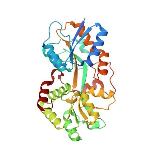
A novel mode of ferric ion coordination by the periplasmic ferric ion-binding subunit FbpA of an ABC-type iron transporter from Thermus thermophilus HB8.

Wang, S.Ogata, M.Horita, S.Ohtsuka, J.Nagata, K.Tanokura, M.

(2014) Acta Crystallogr D Biol Crystallogr 70: 196-202

  • DOI: https://doi.org/10.1107/S1399004713026333
  • Primary Citation of Related Structures:  
    3WAE, 3WAF

  • PubMed Abstract: 

    Crystal structures of FbpA, the periplasmic ferric ion-binding protein of an iron-uptake ABC transporter, from Thermus thermophilus HB8 (TtFbpA) have been solved in apo and ferric ion-bound forms at 1.8 and 1.7 Å resolution, respectively. The latter crystal structure shows that the bound ferric ion forms a novel six-coordinated complex with three tyrosine side chains, two bicarbonates and a water molecule in the metal-binding site. The results of gel-filtration chromatography and dynamic light scattering show that TtFbpA exists as a monomer in solution regardless of ferric ion binding and that TtFbpA adopts a more compact conformation in the ferric ion-bound state than in the apo state in solution.


  • Organizational Affiliation

    Department of Applied Biological Chemistry, Graduate School of Agricultural and Life Sciences, University of Tokyo, 1-1-1 Yayoi, Bunkyo-ku, Tokyo 113-8657, Japan.


Macromolecules
Find similar proteins by:  (by identity cutoff)  |  3D Structure
Entity ID: 1
MoleculeChains Sequence LengthOrganismDetailsImage
IRON ABC TRANSPORTER, PERIPLASMIC IRON-BINDING PROTEIN
A, B
330Thermus thermophilusMutation(s): 0 
Gene Names: TTHA1628
UniProt
Find proteins for Q5SHV2 (Thermus thermophilus (strain ATCC 27634 / DSM 579 / HB8))
Explore Q5SHV2 
Go to UniProtKB:  Q5SHV2
Entity Groups  
Sequence Clusters30% Identity50% Identity70% Identity90% Identity95% Identity100% Identity
UniProt GroupQ5SHV2
Sequence Annotations
Expand
  • Reference Sequence
Experimental Data & Validation

Experimental Data

  • Method: X-RAY DIFFRACTION
  • Resolution: 1.70 Å
  • R-Value Free: 0.243 
  • R-Value Work: 0.201 
  • R-Value Observed: 0.203 
  • Space Group: P 21 21 21
Unit Cell:
Length ( Å )Angle ( ˚ )
a = 40.17α = 90
b = 101.02β = 90
c = 134.61γ = 90
Software Package:
Software NamePurpose
ADSCdata collection
PHASERphasing
REFMACrefinement
XDSMEdata reduction
XDSMEdata scaling

Structure Validation

View Full Validation Report


Entry History 

Deposition Data

Revision History  (Full details and data files)

  • Version 1.0: 2014-05-21
    Type: Initial release
  • Version 1.1: 2024-03-20
    Changes: Data collection, Database references, Derived calculations