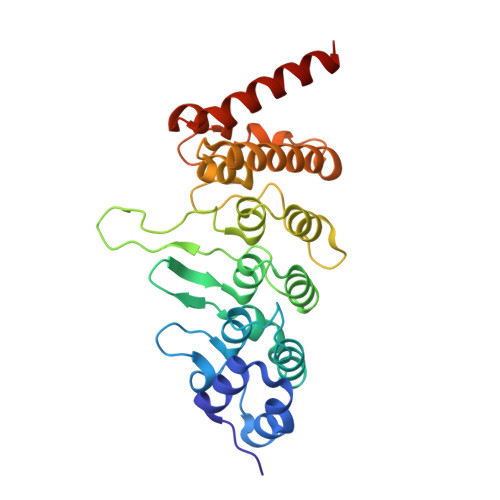
TRPV4 channel activity is modulated by direct interaction of the ankyrin domain to PI(4,5)P2
Takahashi, N., Hamada-Nakahara, S., Itoh, Y., Takemura, K., Shimada, A., Ueda, Y., Kitamata, M., Matsuoka, R., Hanawa-Suetsugu, K., Senju, Y., Mori, M.X., Kiyonaka, S., Kohda, D., Kitao, A., Mori, Y., Suetsugu, S.(2014) Nat Commun 5: 4994-4994
- PubMed: 25256292 Search on PubMed
- DOI: https://doi.org/10.1038/ncomms5994
- Primary Citation Related Structures:
3W9F, 3W9G - PubMed Abstract:
Mutations in the ankyrin repeat domain (ARD) of TRPV4 are responsible for several channelopathies, including Charcot-Marie-Tooth disease type 2C and congenital distal and scapuloperoneal spinal muscular atrophy. However, the molecular pathogenesis mediated by these mutations remains elusive, mainly due to limited understanding of the TRPV4 ARD function. Here we show that phosphoinositide binding to the TRPV4 ARD leads to suppression of the channel activity. Among the phosphoinositides, phosphatidylinositol-4,5-bisphosphate (PI(4,5)P2) most potently binds to the TRPV4 ARD. The crystal structure of the TRPV4 ARD in complex with inositol-1,4,5-trisphosphate, the head-group of PI(4,5)P2, and the molecular-dynamics simulations revealed the PI(4,5)P2-binding amino-acid residues. The TRPV4 channel activities were increased by titration or hydrolysis of membrane PI(4,5)P2. Notably, disease-associated TRPV4 mutations that cause a gain-of-function phenotype abolished PI(4,5)P2 binding and PI(4,5)P2 sensitivity. These findings identify TRPV4 ARD as a lipid-binding domain in which interactions with PI(4,5)P2 normalize the channel activity in TRPV4.
- 1] Laboratory of Molecular Biology, Department of Synthetic Chemistry and Biological Chemistry, Graduate School of Engineering, and Laboratory of Environmental Systems Biology, Department of Technology and Ecology, Hall of Global Environmental Studies, Kyoto University, Kyoto 615-8510, Japan [2] Advanced Biomedical Engineering Research Unit, Kyoto University, Kyoto 615-8510, Japan.
Organizational Affiliation: