3W8I

Crystal structure of CCM3 in complex with the C-terminal regulatory domain of MST4


Experimental Data Snapshot

  • Method: X-RAY DIFFRACTION
  • Resolution: 2.40 Å
  • R-Value Free: 0.267 
  • R-Value Work: 0.222 
  • R-Value Observed: 0.225 

wwPDB Validation   3D Report Full Report


This is version 1.1 of the entry. See complete history


Literature

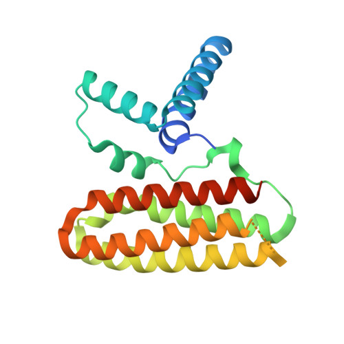
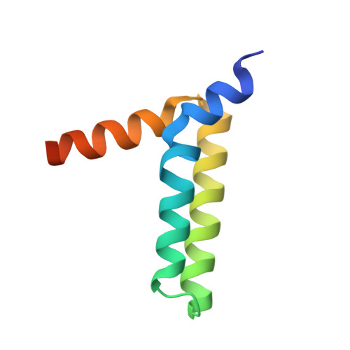
Structural Basis for the Unique Heterodimeric Assembly between Cerebral Cavernous Malformation 3 and Germinal Center Kinase III.

Xu, X.Wang, X.Zhang, Y.Wang, D.C.Ding, J.

(2013) Structure 21: 1059-1066

  • DOI: https://doi.org/10.1016/j.str.2013.04.007
  • Primary Citation of Related Structures:  
    3W8H, 3W8I

  • PubMed Abstract: 

    Defects in cerebral cavernous malformation protein CCM3 result in cerebral cavernous malformation (CCM), a common vascular lesion of the human CNS. CCM3 functions as an adaptor protein that interacts with various signal proteins. Among these partner proteins, germinal center kinase III (GCKIII) proteins have attracted significant interest because GCKIII-CCM3 interactions play essential roles in vascular physiology. Here, we report the crystal structures of CCM3 in complex with the C-terminal regulatory domains of GCKIII (GCKIIIct) at 2.4 Å resolution. Our results reveal that GCKIIIct adopts a fold closely resembling that of the CCM3 N-terminal dimeric domain. GCKIIIct heterodimerizes with CCM3 in a manner analogous to CCM3 homodimerization. The remarkable structural rearrangement of CCM3 induced by GCKIIIct binding and the ensuing interactions within CCM3 are characterized as the structural determinants for GCKIIIct-CCM3 heterodimerization. Taken together, these findings provide a precise structural basis for GCKIIIct-CCM3 heterodimerization and the functional performance of GCKIII mediated by CCM3.


  • Organizational Affiliation

    Institute of Biophysics, Chinese Academy of Sciences, National Laboratory of Biomacromolecules, Beijing 100101, People's Republic of China.


Macromolecules
Find similar proteins by:  (by identity cutoff)  |  3D Structure
Entity ID: 1
MoleculeChains Sequence LengthOrganismDetailsImage
Programmed cell death protein 10213Homo sapiensMutation(s): 0 
Gene Names: CCM3PDCD10TFAR15
UniProt & NIH Common Fund Data Resources
Find proteins for Q9BUL8 (Homo sapiens)
Explore Q9BUL8 
Go to UniProtKB:  Q9BUL8
PHAROS:  Q9BUL8
GTEx:  ENSG00000114209 
Entity Groups  
Sequence Clusters30% Identity50% Identity70% Identity90% Identity95% Identity100% Identity
UniProt GroupQ9BUL8
Sequence Annotations
Expand
  • Reference Sequence
Find similar proteins by:  (by identity cutoff)  |  3D Structure
Entity ID: 2
MoleculeChains Sequence LengthOrganismDetailsImage
Serine/threonine-protein kinase MST476Homo sapiensMutation(s): 0 
Gene Names: MASKMST4SOK1STK25YSK1
EC: 2.7.11.1
UniProt & NIH Common Fund Data Resources
Find proteins for Q9P289 (Homo sapiens)
Explore Q9P289 
Go to UniProtKB:  Q9P289
PHAROS:  Q9P289
GTEx:  ENSG00000134602 
Entity Groups  
Sequence Clusters30% Identity50% Identity70% Identity90% Identity95% Identity100% Identity
UniProt GroupQ9P289
Sequence Annotations
Expand
  • Reference Sequence
Experimental Data & Validation

Experimental Data

  • Method: X-RAY DIFFRACTION
  • Resolution: 2.40 Å
  • R-Value Free: 0.267 
  • R-Value Work: 0.222 
  • R-Value Observed: 0.225 
  • Space Group: P 41 21 2
Unit Cell:
Length ( Å )Angle ( ˚ )
a = 69.1α = 90
b = 69.1β = 90
c = 117.57γ = 90
Software Package:
Software NamePurpose
CrystalCleardata collection
PHASERphasing
PHENIXrefinement
MOSFLMdata reduction
SCALAdata scaling

Structure Validation

View Full Validation Report


Entry History 

Deposition Data

Revision History  (Full details and data files)

  • Version 1.0: 2013-07-03
    Type: Initial release
  • Version 1.1: 2023-11-08
    Changes: Data collection, Database references, Refinement description