3W2T | pdb_00003w2t

Crystal structure of human depiptidyl peptidase IV (DPP-4) in complex with vildagliptin


Experimental Data Snapshot

  • Method: X-RAY DIFFRACTION
  • Resolution: 2.36 Å
  • R-Value Free: 
    0.231 (Depositor), 0.226 (DCC) 
  • R-Value Work: 
    0.180 (Depositor), 0.178 (DCC) 
  • R-Value Observed: 
    0.183 (Depositor) 

Starting Model: experimental
View more details

wwPDB Validation 3D Report Full Report

Validation slider image for 3W2T

Ligand Structure Quality Assessment 


This is version 3.2 of the entry. See complete history

Literature

A comparative study of the binding modes of recently launched dipeptidyl peptidase IV inhibitors in the active site

Nabeno, M.Akahoshi, F.Kishida, H.Miyaguchi, I.Tanaka, Y.Ishii, S.Kadowaki, T.

(2013) Biochem Biophys Res Commun 434: 191-196

  • DOI: https://doi.org/10.1016/j.bbrc.2013.03.010
  • Primary Citation Related Structures: 
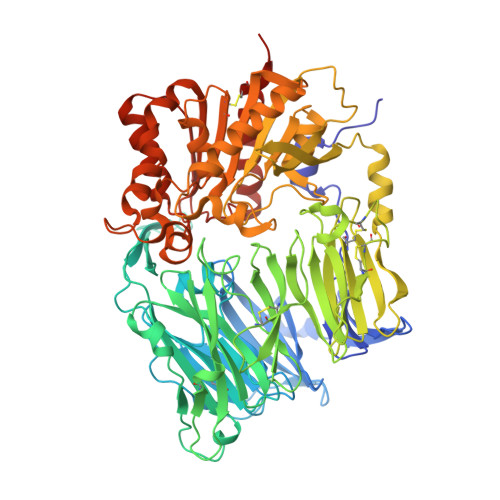
    3W2T

  • PubMed Abstract: 

    In recent years, various dipeptidyl peptidase IV (DPP-4) inhibitors have been released as therapeutic drugs for type 2 diabetes in many countries. In spite of their diverse chemical structures, no comparative studies of their binding modes in the active site of DPP-4 have been disclosed. We determined the co-crystal structure of vildagliptin with DPP-4 by X-ray crystallography and compared the binding modes of six launched inhibitors in DPP-4. The inhibitors were categorized into three classes on the basis of their binding subsites: (i) vildagliptin and saxagliptin (Class 1) form interactions with the core S1 and S2 subsites and a covalent bond with Ser630 in the catalytic triad; (ii) alogliptin and linagliptin (Class 2) form interactions with the S1' and/or S2' subsites in addition to the S1 and S2 subsites; and (iii) sitagliptin and teneligliptin (Class 3) form interactions with the S1, S2 and S2 extensive subsites. The present study revealed that the additional interactions with the S1', S2' or S2 extensive subsite may increase DPP-4 inhibition beyond the level afforded by the fundamental interactions with the S1 and S2 subsites and are more effective than forming a covalent bond with Ser630.


  • Organizational Affiliation
    • Medicinal Chemistry Research Laboratories II, Mitsubishi Tanabe Pharma Corporation, Saitama, Japan. nabeno.mika@mv.mt-pharma.co.jp

Macromolecule Content 

  • Total Structure Weight: 176.94 kDa 
  • Atom Count: 13,311 
  • Modeled Residue Count: 1,458 
  • Deposited Residue Count: 1,480 
  • Unique protein chains: 1

Macromolecules

Find similar proteins by:|  3D Structure
Entity ID: 1
MoleculeChains  Sequence LengthOrganismDetailsImage
Dipeptidyl peptidase 4
A, B
740Homo sapiensMutation(s): 0 
Gene Names: DPP4ADCP2CD26
EC: 3.4.14.5
UniProt & NIH Common Fund Data Resources
Find proteins for P27487 (Homo sapiens)
Explore P27487 
Go to UniProtKB:  P27487
PHAROS:  P27487
GTEx:  ENSG00000197635 
Entity Groups
Sequence Clusters30% Identity50% Identity70% Identity90% Identity95% Identity100% Identity
UniProt GroupP27487
Glycosylation
Glycosylation Sites: 6Go to GlyGen: P27487-1
Sequence Annotations
Expand
Reference Sequence

Oligosaccharides

Help  
Entity ID: 2
MoleculeChains Length2D Diagram GlycosylationD Interactions
beta-L-fucopyranose-(1-6)-2-acetamido-2-deoxy-beta-D-glucopyranose
C, G
2N-Glycosylation
Glycosylation Resources
GlyTouCan: G61843VN
GlyCosmos: G61843VN
GlyGen: G61843VN
Entity ID: 3
MoleculeChains Length2D Diagram GlycosylationD Interactions
2-acetamido-2-deoxy-beta-D-glucopyranose-(1-4)-2-acetamido-2-deoxy-beta-D-glucopyranose
D, E, F, H, I
D, E, F, H, I, J
2N-Glycosylation
Glycosylation Resources
GlyTouCan: G42666HT
GlyCosmos: G42666HT
GlyGen: G42666HT

Small Molecules

Ligands 3 Unique
IDChains Name / Formula / InChI Key2D Diagram3D Interactions
LF7

Query on LF7


Download:Ideal Coordinates CCD File
K [auth A],
P [auth B]
2-{[(1r,3s,5R,7S)-3-hydroxytricyclo[3.3.1.1~3,7~]decan-1-yl]amino}-1-{(2S)-2-[(E)-iminomethyl]pyrrolidin-1-yl}ethan-1-o ne
C17 H27 N3 O2
LUWVSODOPBYGON-FEZPVXCISA-N
NAG

Query on NAG


Download:Ideal Coordinates CCD File
M [auth A],
N [auth A],
O [auth A],
R [auth B],
S [auth B]
2-acetamido-2-deoxy-beta-D-glucopyranose
C8 H15 N O6
OVRNDRQMDRJTHS-FMDGEEDCSA-N
GOL

Query on GOL


Download:Ideal Coordinates CCD File
L [auth A],
Q [auth B]
GLYCEROL
C3 H8 O3
PEDCQBHIVMGVHV-UHFFFAOYSA-N

Experimental Data & Validation

Experimental Data

  • Method: X-RAY DIFFRACTION
  • Resolution: 2.36 Å
  • R-Value Free:  0.231 (Depositor), 0.226 (DCC) 
  • R-Value Work:  0.180 (Depositor), 0.178 (DCC) 
  • R-Value Observed: 0.183 (Depositor) 
Space Group: P 21 21 21
Unit Cell:
Length ( Å )Angle ( ˚ )
a = 118.216α = 90
b = 126.237β = 90
c = 137.089γ = 90
Software Package:
Software NamePurpose
ADSCdata collection
PHASERphasing
REFMACrefinement
HKL-2000data reduction
SCALEPACKdata scaling

Structure Validation

View Full Validation Report


Ligand Structure Quality Assessment 


Entry History 

Deposition Data

Revision History  (Full details and data files)

  • Version 1.0: 2013-05-15
    Type: Initial release
  • Version 2.0: 2018-02-14
    Changes: Non-polymer description, Structure summary
  • Version 3.0: 2020-07-29
    Type: Remediation
    Reason: Carbohydrate remediation
    Changes: Atomic model, Data collection, Database references, Derived calculations, Structure summary
  • Version 3.1: 2023-11-08
    Changes: Data collection, Database references, Refinement description, Structure summary
  • Version 3.2: 2024-10-30
    Changes: Structure summary