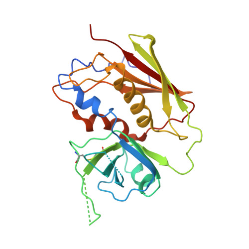
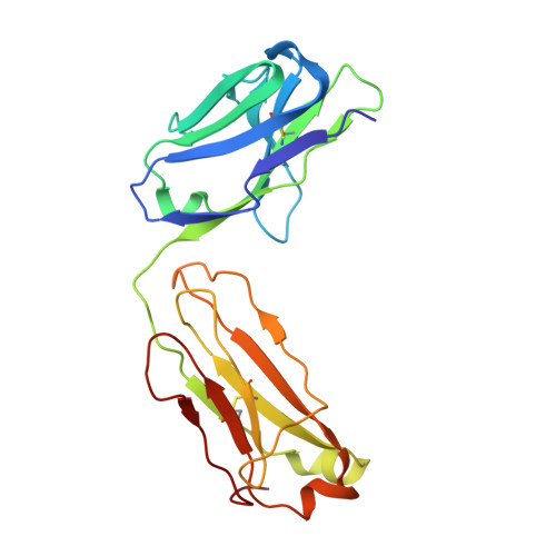
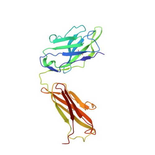
3W2D

Crystal Structure of Staphylococcal Eenterotoxin B in complex with a novel neutralization monoclonal antibody Fab fragment


Experimental Data Snapshot

  • Method: X-RAY DIFFRACTION
  • Resolution: 3.10 Å
  • R-Value Free: 0.261 
  • R-Value Work: 0.196 
  • R-Value Observed: 0.200 

wwPDB Validation   3D Report Full Report


This is version 1.1 of the entry. See complete history


Literature

Structural basis for the neutralization and specificity of Staphylococcal enterotoxin B against its MHC Class II binding site.

Xia, T.Liang, S.Wang, H.Hu, S.Sun, Y.Yu, X.Han, J.Li, J.Guo, S.Dai, J.Lou, Z.Guo, Y.

(2014) MAbs 6: 119-129

  • DOI: https://doi.org/10.4161/mabs.27106
  • Primary Citation of Related Structures:  
    3W2D

  • PubMed Abstract: 

    Staphylococcal enterotoxin (SE) B is among the potent toxins produced by Staphylococcus aureus that cause toxic shock syndrome (TSS), which can result in multi-organ failure and death. Currently, neutralizing antibodies have been shown to be effective immunotherapeutic agents against this toxin, but the structural basis of the neutralizing mechanism is still unknown. In this study, we generated a neutralizing monoclonal antibody, 3E2, against SEB, and analyzed the crystal structure of the SEB-3E2 Fab complex. Crystallographic analysis suggested that the neutralizing epitope overlapped with the MHC II molecule binding site on SEB, and thus 3E2 could inhibit SEB function by preventing interaction with the MHC II molecule. Mutagenesis studies were done on SEB, as well as the related Staphylococcus aureus toxins SEA and SEC. These studies revealed that tyrosine (Y)46 and lysine (K)71 residues of SEB are essential to specific antibody-antigen recognition and neutralization. Substitution of Y at SEA glutamine (Q)49, which corresponds to SEB Y46, increased both 3E2's binding to SEA in vitro and the neutralization of SEA in vivo. These results suggested that SEB Y46 is responsible for distinguishing SEB from SEA. These findings may be helpful for the development of antibody-based therapy for SEB-induced TSS.


  • Organizational Affiliation

    International Joint Cancer Institute; Second Military Medical University; Shanghai, P.R. China; College of Pharmacy; Liaocheng University; Liaocheng, P.R. China.


Macromolecules
Find similar proteins by:  (by identity cutoff)  |  3D Structure
Entity ID: 1
MoleculeChains Sequence LengthOrganismDetailsImage
Enterotoxin type B239Staphylococcus aureusMutation(s): 0 
Gene Names: entB
UniProt
Find proteins for P01552 (Staphylococcus aureus)
Explore P01552 
Go to UniProtKB:  P01552
Entity Groups  
Sequence Clusters30% Identity50% Identity70% Identity90% Identity95% Identity100% Identity
UniProt GroupP01552
Sequence Annotations
Expand
  • Reference Sequence
Find similar proteins by:  (by identity cutoff)  |  3D Structure
Entity ID: 2
MoleculeChains Sequence LengthOrganismDetailsImage
Monoclonal Antibody 3E2 Fab figment light chainB [auth L]215Mus musculusMutation(s): 0 
Entity Groups  
Sequence Clusters30% Identity50% Identity70% Identity90% Identity95% Identity100% Identity
Sequence Annotations
Expand
  • Reference Sequence
Find similar proteins by:  (by identity cutoff)  |  3D Structure
Entity ID: 3
MoleculeChains Sequence LengthOrganismDetailsImage
Monoclonal Antibody 3E2 Fab figment heavy chainC [auth H]223Mus musculusMutation(s): 0 
Entity Groups  
Sequence Clusters30% Identity50% Identity70% Identity90% Identity95% Identity100% Identity
Sequence Annotations
Expand
  • Reference Sequence
Small Molecules
Ligands 1 Unique
IDChains Name / Formula / InChI Key2D Diagram3D Interactions
SO4
Query on SO4

Download Ideal Coordinates CCD File 
D [auth H]SULFATE ION
O4 S
QAOWNCQODCNURD-UHFFFAOYSA-L
Experimental Data & Validation

Experimental Data

  • Method: X-RAY DIFFRACTION
  • Resolution: 3.10 Å
  • R-Value Free: 0.261 
  • R-Value Work: 0.196 
  • R-Value Observed: 0.200 
  • Space Group: P 1 2 1
Unit Cell:
Length ( Å )Angle ( ˚ )
a = 40.561α = 90
b = 109.308β = 96.08
c = 86.222γ = 90
Software Package:
Software NamePurpose
PHENIXrefinement

Structure Validation

View Full Validation Report


Entry History 

Deposition Data

Revision History  (Full details and data files)

  • Version 1.0: 2013-12-25
    Type: Initial release
  • Version 1.1: 2014-04-16
    Changes: Database references, Source and taxonomy