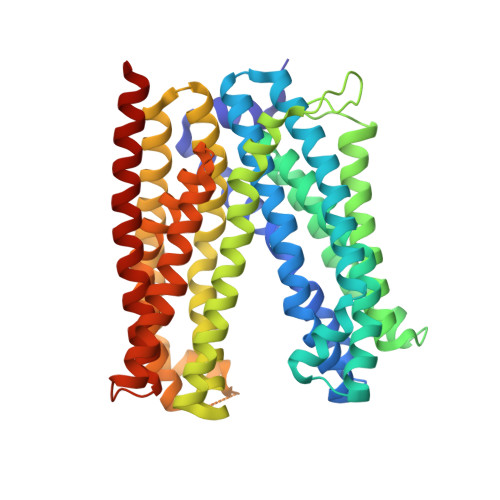
Structural basis for the drug extrusion mechanism by a MATE multidrug transporter.
Tanaka, Y., Hipolito, C.J., Maturana, A.D., Ito, K., Kuroda, T., Higuchi, T., Katoh, T., Kato, H.E., Hattori, M., Kumazaki, K., Tsukazaki, T., Ishitani, R., Suga, H., Nureki, O.(2013) Nature 496: 247-251
- PubMed: 23535598
- DOI: https://doi.org/10.1038/nature12014
- Primary Citation of Related Structures:
3VVN, 3VVO, 3VVP, 3VVR, 3VVS, 3W4T, 3WBN - PubMed Abstract:
Multidrug and toxic compound extrusion (MATE) family transporters are conserved in the three primary domains of life (Archaea, Bacteria and Eukarya), and export xenobiotics using an electrochemical gradient of H(+) or Na(+) across the membrane. MATE transporters confer multidrug resistance to bacterial pathogens and cancer cells, thus causing critical reductions in the therapeutic efficacies of antibiotics and anti-cancer drugs, respectively. Therefore, the development of MATE inhibitors has long been awaited in the field of clinical medicine. Here we present the crystal structures of the H(+)-driven MATE transporter from Pyrococcus furiosus in two distinct apo-form conformations, and in complexes with a derivative of the antibacterial drug norfloxacin and three in vitro selected thioether-macrocyclic peptides, at 2.1-3.0 Å resolutions. The structures, combined with functional analyses, show that the protonation of Asp 41 on the amino (N)-terminal lobe induces the bending of TM1, which in turn collapses the N-lobe cavity, thereby extruding the substrate drug to the extracellular space. Moreover, the macrocyclic peptides bind the central cleft in distinct manners, which correlate with their inhibitory activities. The strongest inhibitory peptide that occupies the N-lobe cavity may pave the way towards the development of efficient inhibitors against MATE transporters.
- RIKEN Advanced Science Institute, 2-1 Hirosawa, Wako-shi, Saitama 351-0198, Japan.
Organizational Affiliation:


