


X-ray structure of the human mitogen-activated protein kinase kinase 1 (MEK1) in complex with an inhibitor and MgATP
Kudo, N., Kato, R., Wakatsuki, S.To be published.
Experimental Data Snapshot
Starting Model: experimental
View more details
Entity ID: 1 | |||||
|---|---|---|---|---|---|
| Molecule | Chains | Sequence Length | Organism | Details | Image |
| Dual specificity mitogen-activated protein kinase kinase 1 | 341 | Homo sapiens | Mutation(s): 0 Gene Names: MAP2K1, MEK1, PRKMK1 EC: 2.7.12.2 | 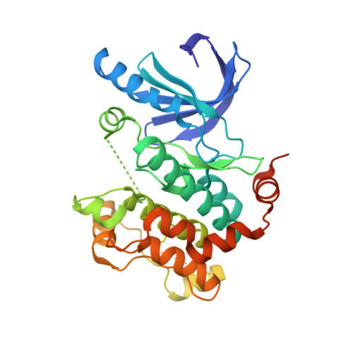 | |
UniProt & NIH Common Fund Data Resources | |||||
PHAROS: Q02750 GTEx: ENSG00000169032 | |||||
Entity Groups | |||||
| Sequence Clusters | 30% Identity50% Identity70% Identity90% Identity95% Identity100% Identity | ||||
| UniProt Group | Q02750 | ||||
Sequence AnnotationsExpand | |||||
Reference Sequence | |||||
| Ligands 3 Unique | |||||
|---|---|---|---|---|---|
| ID | Chains | Name / Formula / InChI Key | 2D Diagram | 3D Interactions | |
| ATP Download:Ideal Coordinates CCD File | E [auth A], H [auth B], K [auth C] | ADENOSINE-5'-TRIPHOSPHATE C10 H16 N5 O13 P3 ZKHQWZAMYRWXGA-KQYNXXCUSA-N |  | ||
| 4BM Download:Ideal Coordinates CCD File | F [auth A], I [auth B], L [auth C] | N-{[(2R)-2,3-dihydroxypropyl]oxy}-3,4-difluoro-2-[(2-fluoro-4-iodophenyl)amino]benzamide C16 H14 F3 I N2 O4 SUDAHWBOROXANE-SECBINFHSA-N |  | ||
| MG Download:Ideal Coordinates CCD File | D [auth A], G [auth B], J [auth C] | MAGNESIUM ION Mg JLVVSXFLKOJNIY-UHFFFAOYSA-N |  | ||
| Length ( Å ) | Angle ( ˚ ) |
|---|---|
| a = 58.894 | α = 90 |
| b = 129.09 | β = 90 |
| c = 135.736 | γ = 90 |
| Software Name | Purpose |
|---|---|
| HKL-2000 | data collection |
| MOLREP | phasing |
| REFMAC | refinement |
| HKL-2000 | data reduction |
| HKL-2000 | data scaling |


