Small-angle X-ray scattering reveals structural dynamics of the botulinum neurotoxin associating protein, nontoxic nonhemagglutinin
Sagane, Y., Miyashita, S., Miyata, K., Matsumoto, T., Inui, K., Hayashi, S., Suzuki, T., Hasegawa, K., Yajima, S., Yamano, A., Niwa, K., Watanabe, T.(2012) Biochem Biophys Res Commun 425: 256-260
- PubMed: 22828508 Search on PubMed
- DOI: https://doi.org/10.1016/j.bbrc.2012.07.077
- Primary Citation Related Structures:
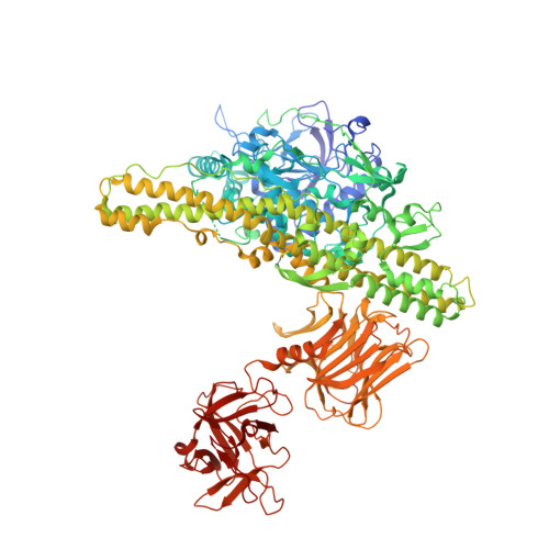
3VUO - PubMed Abstract:
In cell culture supernatants, the botulinum neurotoxin (BoNT) exists as part of a toxin complex (TC) in which nontoxic nonhemagglutinin (NTNHA) and/or hemagglutinins (HAs) are assembled onto the BoNT. A series of investigations indicated that formation of the TC is vital for delivery of the toxin to nerve cells through the digestive tract. In the assembly process, BoNT binds to NTNHA yielding M-TC, and it then matures into L-TC by further association with the HAs via NTNHA in the M-TC. Here, we report a crystal structure of the NTNHA from Clostridium botulinum serotype D strain 4947. Additionally, we performed small-angle X-ray scattering (SAXS) analysis of the NTNHA and the M-TC to elucidate the solution structure. The crystal structure of D-4947 NTNHA revealed that BoNT and NTNHA share a closely related structure consisting of three domains. The SAXS image indicated that, even though the N-terminal two-thirds of the NTNHA molecule had an apparently similar conformation in both the crystal and solution structures, the C-terminal third of the molecule showed a more extended structure in the SAXS image than that seen in the crystallographic image. The discrepancy between the crystal and solution structures implies a high flexibility of the C-terminal third domain of NTNHA, which is involved in binding to BoNT. Structural dynamics of the NTNHA molecule revealed by SAXS may explain its binding to BoNT to form the BoNT/NTNHA complex.
- Department of Food and Cosmetic Science, Faculty of Bioindustry, Tokyo University of Agriculture, 196 Yasaka, Abashiri 099-2493, Japan.
Organizational Affiliation: