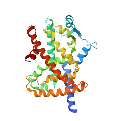
3VSO

Human PPAR gamma ligand binding domain in complex with a gamma selective agonist mekt21


Experimental Data Snapshot

  • Method: X-RAY DIFFRACTION
  • Resolution: 2.00 Å
  • R-Value Free: 0.257 
  • R-Value Work: 0.219 
  • R-Value Observed: 0.221 

Starting Model: experimental
View more details

wwPDB Validation   3D Report Full Report


Ligand Structure Quality Assessment 


This is version 1.1 of the entry. See complete history


Literature

Design and synthesis of a series of alpha-benzyl phenylpropanoic acid-type peroxisome proliferator-activated receptor (PPAR) gamma partial agonists with improved aqueous solubility

Ohashi, M.Oyama, T.Putranto, E.W.Waku, T.Nobusada, H.Kataoka, K.Matsuno, K.Yashiro, M.Morikawa, K.Huh, N.H.Miyachi, H.

(2013) Bioorg Med Chem 21: 2319-2332

  • DOI: https://doi.org/10.1016/j.bmc.2013.02.003
  • Primary Citation of Related Structures:  
    3VSO

  • PubMed Abstract: 

    In the continuing study directed toward the development of peroxisome proliferator-activated receptor gamma (hPPARγ) agonist, we attempted to improve the water solubility of our previously developed hPPARγ-selective agonist 3, which is insufficiently soluble for practical use, by employing two strategies: introducing substituents to reduce its molecular planarity and decreasing its hydrophobicity via replacement of the adamantyl group with a heteroaromatic ring. The first approach proved ineffective, but the second was productive. Here, we report the design and synthesis of a series of α-benzyl phenylpropanoic acid-type hPPARγ partial agonists with improved aqueous solubility. Among them, we selected (R)-7j, which activates hPPARγ to the extent of about 65% of the maximum observed with a full agonist, for further evaluation. The ligand-binding mode and the reason for the partial-agonistic activity are discussed based on X-ray-determined structure of the complex of hPPARγ ligand-binding domain (LBD) and (R)-7j with previously reported ligand-LDB structures. Preliminal apoptotic effect of (R)-7j against human scirrhous gastric cancer cell line OCUM-2MD3 is also described.


  • Organizational Affiliation

    Graduate School of Medicine, Dentistry and Pharmaceutical Sciences, Okayama University, 1-1-1 Tsushima-Naka, Kita-ku, Okayama 700-8530, Japan.


Macromolecules
Find similar proteins by:  (by identity cutoff)  |  3D Structure
Entity ID: 1
MoleculeChains Sequence LengthOrganismDetailsImage
Peroxisome proliferator-activated receptor gamma
A, B
286Homo sapiensMutation(s): 0 
Gene Names: PPARGNR1C3
UniProt & NIH Common Fund Data Resources
Find proteins for P37231 (Homo sapiens)
Explore P37231 
Go to UniProtKB:  P37231
PHAROS:  P37231
GTEx:  ENSG00000132170 
Entity Groups  
Sequence Clusters30% Identity50% Identity70% Identity90% Identity95% Identity100% Identity
UniProt GroupP37231
Sequence Annotations
Expand
  • Reference Sequence
Small Molecules
Ligands 1 Unique
IDChains Name / Formula / InChI Key2D Diagram3D Interactions
EK1
Query on EK1

Download Ideal Coordinates CCD File 
C [auth A](2R)-2-benzyl-3-[4-propoxy-3-({[4-(pyrimidin-2-yl)benzoyl]amino}methyl)phenyl]propanoic acid
C31 H31 N3 O4
DEAVJKJURZRSJV-AREMUKBSSA-N
Binding Affinity Annotations 
IDSourceBinding Affinity
EK1 BindingDB:  3VSO EC50: 35 (nM) from 1 assay(s)
Experimental Data & Validation

Experimental Data

  • Method: X-RAY DIFFRACTION
  • Resolution: 2.00 Å
  • R-Value Free: 0.257 
  • R-Value Work: 0.219 
  • R-Value Observed: 0.221 
  • Space Group: C 1 2 1
Unit Cell:
Length ( Å )Angle ( ˚ )
a = 93.346α = 90
b = 61.706β = 102.79
c = 118.984γ = 90
Software Package:
Software NamePurpose
PHENIXrefinement

Structure Validation

View Full Validation Report


Ligand Structure Quality Assessment 


Entry History 

Deposition Data

Revision History  (Full details and data files)

  • Version 1.0: 2013-05-01
    Type: Initial release
  • Version 1.1: 2023-11-08
    Changes: Data collection, Database references, Derived calculations, Refinement description