3VRJ | pdb_00003vrj

HLA-B*57:01-LTTKLTNTNI in complex with abacavir


Experimental Data Snapshot

  • Method: X-RAY DIFFRACTION
  • Resolution: 1.90 Å
  • R-Value Free: 
    0.213 (Depositor), 0.212 (DCC) 
  • R-Value Work: 
    0.175 (Depositor), 0.174 (DCC) 
  • R-Value Observed: 
    0.177 (Depositor) 

Starting Model: experimental
View more details

wwPDB Validation 3D Report Full Report

Validation slider image for 3VRJ

Ligand Structure Quality Assessment 


This is version 1.3 of the entry. See complete history

Literature

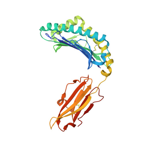
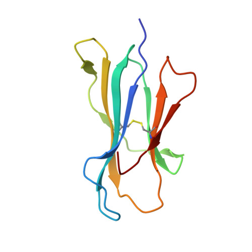
Immune self-reactivity triggered by drug-modified HLA-peptide repertoire

Illing, P.T.Vivian, J.P.Dudek, N.L.Kostenko, L.Chen, Z.Bharadwaj, M.Miles, J.J.Kjer-Nielsen, L.Gras, S.Williamson, N.A.Burrows, S.R.Purcell, A.W.Rossjohn, J.McCluskey, J.

(2012) Nature 486: 554-558

  • DOI: https://doi.org/10.1038/nature11147
  • Primary Citation Related Structures: 
    3VRI, 3VRJ

  • PubMed Abstract: 

    Human leukocyte antigens (HLAs) are highly polymorphic proteins that initiate immunity by presenting pathogen-derived peptides to T cells. HLA polymorphisms mostly map to the antigen-binding cleft, thereby diversifying the repertoire of self-derived and pathogen-derived peptide antigens selected by different HLA allotypes. A growing number of immunologically based drug reactions, including abacavir hypersensitivity syndrome (AHS) and carbamazepine-induced Stevens-Johnson syndrome (SJS), are associated with specific HLA alleles. However, little is known about the underlying mechanisms of these associations, including AHS, a prototypical HLA-associated drug reaction occurring exclusively in individuals with the common histocompatibility allele HLA-B*57:01, and with a relative risk of more than 1,000 (refs 6, 7). We show that unmodified abacavir binds non-covalently to HLA-B*57:01, lying across the bottom of the antigen-binding cleft and reaching into the F-pocket, where a carboxy-terminal tryptophan typically anchors peptides bound to HLA-B*57:01. Abacavir binds with exquisite specificity to HLA-B*57:01, changing the shape and chemistry of the antigen-binding cleft, thereby altering the repertoire of endogenous peptides that can bind HLA-B*57:01. In this way, abacavir guides the selection of new endogenous peptides, inducing a marked alteration in 'immunological self'. The resultant peptide-centric 'altered self' activates abacavir-specific T-cells, thereby driving polyclonal CD8 T-cell activation and a systemic reaction manifesting as AHS. We also show that carbamazepine, a widely used anti-epileptic drug associated with hypersensitivity reactions in HLA-B*15:02 individuals, binds to this allotype, producing alterations in the repertoire of presented self peptides. Our findings simultaneously highlight the importance of HLA polymorphism in the evolution of pharmacogenomics and provide a general mechanism for some of the growing number of HLA-linked hypersensitivities that involve small-molecule drugs.


  • Organizational Affiliation
    • Department of Microbiology & Immunology, University of Melbourne, Parkville, Victoria 3010, Australia.

Macromolecule Content 

  • Total Structure Weight: 45.02 kDa 
  • Atom Count: 3,609 
  • Modeled Residue Count: 384 
  • Deposited Residue Count: 386 
  • Unique protein chains: 3

Macromolecules

Find similar proteins by:|  3D Structure
Entity ID: 1
MoleculeChains  Sequence LengthOrganismDetailsImage
HLA class I histocompatibility antigen, B-57 alpha chain276Homo sapiensMutation(s): 0 
Gene Names: HLA-B
UniProt & NIH Common Fund Data Resources
Find proteins for P01889 (Homo sapiens)
Explore P01889 
Go to UniProtKB:  P01889
PHAROS:  P01889
GTEx:  ENSG00000234745 
Entity Groups
Sequence Clusters30% Identity50% Identity70% Identity90% Identity95% Identity100% Identity
UniProt GroupP01889
Sequence Annotations
Expand
Reference Sequence
Find similar proteins by:|  3D Structure
Entity ID: 2
MoleculeChains  Sequence LengthOrganismDetailsImage
Beta-2-microglobulin100Homo sapiensMutation(s): 0 
Gene Names: B2M
UniProt & NIH Common Fund Data Resources
Find proteins for P61769 (Homo sapiens)
Explore P61769 
Go to UniProtKB:  P61769
PHAROS:  P61769
GTEx:  ENSG00000166710 
Entity Groups
Sequence Clusters30% Identity50% Identity70% Identity90% Identity95% Identity100% Identity
UniProt GroupP61769
Sequence Annotations
Expand
Reference Sequence
Find similar proteins by:  Sequence   |   3D Structure  
Entity ID: 3
MoleculeChains  Sequence LengthOrganismDetailsImage
10-mer peptide10N/AMutation(s): 0 
EC: 7.1.1.9
UniProt & NIH Common Fund Data Resources
Find proteins for P00403 (Homo sapiens)
Explore P00403 
Go to UniProtKB:  P00403
PHAROS:  P00403
GTEx:  ENSG00000198712 
Entity Groups
Sequence Clusters30% Identity50% Identity70% Identity90% Identity95% Identity100% Identity
UniProt GroupP00403
Sequence Annotations
Expand
Reference Sequence

Small Molecules

Ligands 1 Unique
IDChains Name / Formula / InChI Key2D Diagram3D Interactions
1KX

Query on 1KX


Download:Ideal Coordinates CCD File
D [auth A]{(1S,4R)-4-[2-amino-6-(cyclopropylamino)-9H-purin-9-yl]cyclopent-2-en-1-yl}methanol
C14 H18 N6 O
MCGSCOLBFJQGHM-SCZZXKLOSA-N

Experimental Data & Validation

Experimental Data

  • Method: X-RAY DIFFRACTION
  • Resolution: 1.90 Å
  • R-Value Free:  0.213 (Depositor), 0.212 (DCC) 
  • R-Value Work:  0.175 (Depositor), 0.174 (DCC) 
  • R-Value Observed: 0.177 (Depositor) 
Space Group: P 1 21 1
Unit Cell:
Length ( Å )Angle ( ˚ )
a = 64.291α = 90
b = 49.005β = 99.53
c = 70.875γ = 90
Software Package:
Software NamePurpose
Blu-Icedata collection
PHASERphasing
PHENIXrefinement
MOSFLMdata reduction
SCALAdata scaling

Structure Validation

View Full Validation Report


Ligand Structure Quality Assessment 


Entry History 

Deposition Data

Revision History  (Full details and data files)

  • Version 1.0: 2012-05-30
    Type: Initial release
  • Version 1.1: 2012-07-04
    Changes: Database references
  • Version 1.2: 2023-11-08
    Changes: Data collection, Database references, Derived calculations, Refinement description
  • Version 1.3: 2024-11-06
    Changes: Structure summary