Structural basis of the strict phospholipid binding specificity of the pleckstrin homology domain of human evectin-2
Okazaki, S., Kato, R., Uchida, Y., Taguchi, T., Arai, H., Wakatsuki, S.(2012) Acta Crystallogr D Biol Crystallogr 68: 117-123
- PubMed: 22281740 Search on PubMed
- DOI: https://doi.org/10.1107/S0907444911051626
- Primary Citation Related Structures:
3VIA - PubMed Abstract:
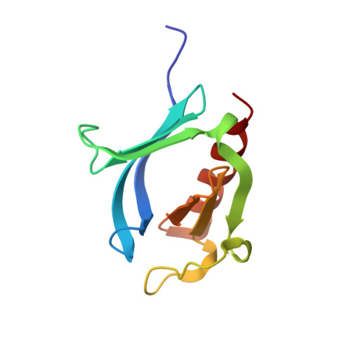
Evectin-2 is a recycling endosomal protein involved in retrograde transport. Its primary sequence contains an N-terminal pleckstrin homology (PH) domain and a C-terminal hydrophobic region. The PH domain of evectin-2 can specifically bind phosphatidylserine, which is enriched in recycling endosomes, and plays an essential role in retrograde transport from recycling endosomes to the trans-Golgi network. The structure of human evectin-2 PH domain in complex with O-phospho-L-serine has recently been reported and demonstrates how the head group of phosphatidylserine is recognized. However, it was not possible to elucidate from the structure why evectin-2 cannot bind phosphatidic acid or phosphatidylethanolamine, which share a common moiety with phosphatidylserine. Here, the crystal structure at 1.75 Å resolution of an apo form of human evectin-2 PH domain, in which the ligand-binding site is free from crystal packing and is thus appropriate for comparison with the structure of the complex, is reported. Comparison between the structures of the apo form and the O-phospho-L-serine complex revealed ligand-induced conformational change evoked by interaction between the carboxyl moiety of the head group of phosphatidylserine and the main-chain N atom of Thr14. This structural change effectively explains the strict ligand specificity of the PH domain of human evectin-2.
- Structural Biology Research Center, Photon Factory, Institute of Materials Structure Science, High Energy Accelerator Research Organization (KEK), Tsukuba, Ibaraki 305-0801, Japan.
Organizational Affiliation: