Design, synthesis, and evaluation of 5-methyl-4-phenoxy-5H-pyrrolo[3,2-d]pyrimidine derivatives: novel VEGFR2 kinase inhibitors binding to inactive kinase conformation.
Oguro, Y., Miyamoto, N., Okada, K., Takagi, T., Iwata, H., Awazu, Y., Miki, H., Hori, A., Kamiyama, K., Imamura, S.(2010) Bioorg Med Chem 18: 7260-7273
- PubMed: 20833055 Search on PubMed
- DOI: https://doi.org/10.1016/j.bmc.2010.08.017
- Primary Citation Related Structures:
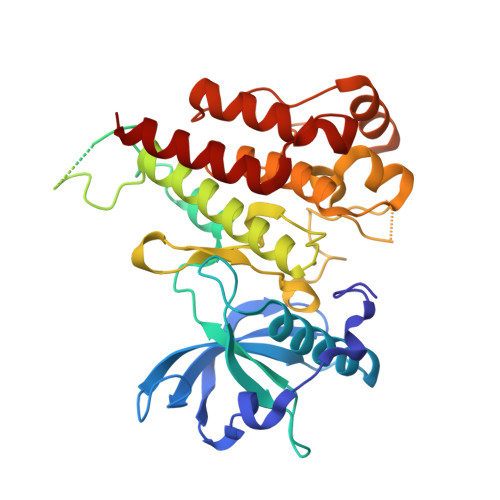
3VHE - PubMed Abstract:
We synthesized a series of pyrrolo[3,2-d]pyrimidine derivatives and evaluated their application as type-II inhibitors of vascular endothelial growth factor receptor 2 (VEGFR2) kinase. Incorporation of a diphenylurea moiety at the C4-position of the pyrrolo[3,2-d]pyrimidine core via an oxygen linker resulted in compounds that were potent inhibitors of VEGFR2 kinase. Of these derivatives, compound 20d showed the strongest inhibition of VEGF-stimulated proliferation of human umbilical vein endothelial cells (HUVEC). The co-crystal structure of 20d and VEGFR2 revealed that 20d binds to the inactive form of VEGFR2. Further studies indicated that 20d inhibited VEGFR2 kinase with slow dissociation kinetics and also inhibited PDGFR and Tie-2 kinases. Oral administration of the hydrochloride salt of 20d at 3mg/kg/day showed potent inhibition of tumor growth in a DU145 human prostate cancer cell xenograft nude mouse model.
- Pharmaceutical Research Division, Takeda Pharmaceutical Co., Ltd, 10, Wadai, Tsukuba, Ibaraki 300-4293, Japan. Ooguro_Yuuya@takeda.co.jp
Organizational Affiliation: