3VC3 | pdb_00003vc3

Crystal structure of beta-cyanoalanine synthase K95A mutant in soybean


Experimental Data Snapshot

  • Method: X-RAY DIFFRACTION
  • Resolution: 1.77 Å
  • R-Value Free: 
    0.202 (Depositor), 0.199 (DCC) 
  • R-Value Work: 
    0.170 (Depositor), 0.170 (DCC) 
  • R-Value Observed: 
    0.170 (Depositor) 

wwPDB Validation 3D Report Full Report

Validation slider image for 3VC3

Ligand Structure Quality Assessment 


This is version 1.1 of the entry. See complete history

Literature

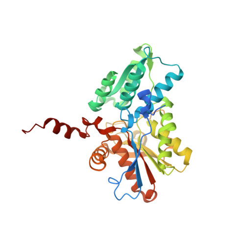
Structure of Soybean beta-Cyanoalanine Synthase and the Molecular Basis for Cyanide Detoxification in Plants.

Yi, H.Juergens, M.Jez, J.M.

(2012) Plant Cell 24: 2696-2706

  • DOI: https://doi.org/10.1105/tpc.112.098954
  • Primary Citation Related Structures: 
    3VBE, 3VC3

  • PubMed Abstract: 

    Plants produce cyanide (CN-) during ethylene biosynthesis in the mitochondria and require β-cyanoalanine synthase (CAS) for CN- detoxification. Recent studies show that CAS is a member of the β-substituted alanine synthase (BSAS) family, which also includes the Cys biosynthesis enzyme O-acetylserine sulfhydrylase (OASS), but how the BSAS evolved distinct metabolic functions is not understood. Here we show that soybean (Glycine max) CAS and OASS form α-aminoacrylate reaction intermediates from Cys and O-acetylserine, respectively. To understand the molecular evolution of CAS and OASS in the BSAS enzyme family, the crystal structures of Gm-CAS and the Gm-CAS K95A mutant with a linked pyridoxal phosphate (PLP)-Cys molecule in the active site were determined. These structures establish a common fold for the plant BSAS family and reveal a substrate-induced conformational change that encloses the active site for catalysis. Comparison of CAS and OASS identified residues that covary in the PLP binding site. The Gm-OASS T81M, S181M, and T185S mutants altered the ratio of OASS:CAS activity but did not convert substrate preference to that of a CAS. Generation of a triple mutant Gm-OASS successfully switched reaction chemistry to that of a CAS. This study provides new molecular insight into the evolution of diverse enzyme functions across the BSAS family in plants.


  • Organizational Affiliation
    • Department of Biology, Washington University, St. Louis, Missouri 63130, USA.

Macromolecule Content 

  • Total Structure Weight: 225.18 kDa 
  • Atom Count: 16,613 
  • Modeled Residue Count: 1,934 
  • Deposited Residue Count: 2,064 
  • Unique protein chains: 1

Macromolecules

Find similar proteins by:|  3D Structure
Entity ID: 1
MoleculeChains  Sequence LengthOrganismDetailsImage
beta-cyanoalnine synthase
A, B, C, D, E
A, B, C, D, E, F
344Glycine maxMutation(s): 0 
Gene Names: GLYMA09G39390OAS-TL3
EC: 2.5.1.47
UniProt
Find proteins for I1L6I6 (Glycine max)
Explore I1L6I6 
Go to UniProtKB:  I1L6I6
Entity Groups
Sequence Clusters30% Identity50% Identity70% Identity90% Identity95% Identity100% Identity
UniProt GroupI1L6I6
Sequence Annotations
Expand
Reference Sequence

Experimental Data & Validation

Experimental Data

  • Method: X-RAY DIFFRACTION
  • Resolution: 1.77 Å
  • R-Value Free:  0.202 (Depositor), 0.199 (DCC) 
  • R-Value Work:  0.170 (Depositor), 0.170 (DCC) 
  • R-Value Observed: 0.170 (Depositor) 
Space Group: P 2 21 21
Unit Cell:
Length ( Å )Angle ( ˚ )
a = 71.092α = 90
b = 154.325β = 90
c = 198.328γ = 90
Software Package:
Software NamePurpose
SCALEPACKdata scaling
PHENIXrefinement
PDB_EXTRACTdata extraction
HKL-3000data collection

Structure Validation

View Full Validation Report


Ligand Structure Quality Assessment 


Entry History 

Deposition Data

Revision History  (Full details and data files)

  • Version 1.0: 2012-09-12
    Type: Initial release
  • Version 1.1: 2024-02-28
    Changes: Data collection, Database references, Derived calculations