Structural Studies of AntD: An N-Acyltransferase Involved in the Biosynthesis of d-Anthrose.
Kubiak, R.L., Holden, H.M.(2012) Biochemistry 51: 867-878
- PubMed: 22220494 Search on PubMed
- DOI: https://doi.org/10.1021/bi201650c
- Primary Citation Related Structures:
3VBI, 3VBJ, 3VBK, 3VBL, 3VBM, 3VBN, 3VBP - PubMed Abstract:
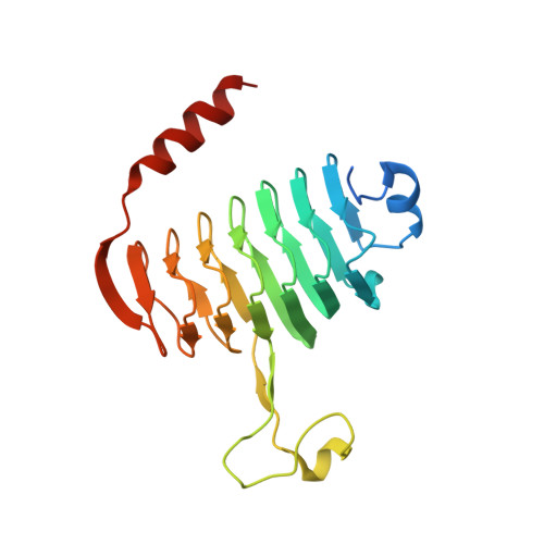
The unusual dideoxy sugar d-anthrose has been identified as an important component in the endospores of infectious agents such as Bacillus anthracis and Bacillus cereus. Specifically, it is the terminal sugar on the bacterium's exosporium, and it provides a point of interaction between the spore and the host. The biosynthesis of d-anthrose involves numerous steps starting from α-d-glucose 1-phosphate. Here we present a combined structural and functional investigation of AntD from B. cereus. This enzyme plays a key role in d-anthrose biosynthesis by catalyzing the acylation of the C-4″ amino group of dTDP-4-amino-4,6-dideoxyglucose using 3-hydroxy-3-methylbutyryl-CoA as the acyl donor. For this investigation, two ternary complexes of AntD were determined to 1.8 Å resolution: one in which the protein contained bound β-hydroxybutyryl-CoA and dTDP and the second with CoA and dTDP-4-amino-4,6-dideoxyglucose. On the basis of these high-resolution structures, it was shown that the side chain of Asp 94 lies within hydrogen bonding distance of the sugar C-4″ amino group, and the side chain of Ser 84 resides near the carbonyl oxygen of β-hydroxybutyryl-CoA. To test the roles of these residues in the catalytic mechanism of AntD, various site-directed mutant proteins were prepared and subjected to kinetic and structural analyses. The D94A and D94N mutant proteins demonstrated enzymatic activity, albeit with significantly reduced catalytic efficiencies. The S84A mutant protein showed an approximate 10-fold decrease in activity. Interestingly, the S84C and S84T mutant proteins were both active but demonstrated substrate inhibition. The three-dimensional structures of all of the mutant proteins were nearly identical to that of the wild-type enzyme, indicating that the changes in their kinetic parameters were not due to major conformational changes. Taken together, these data suggest that Asp 94 is important for substrate binding, but probably does not function as an enzymatic base, and that Ser 84 most likely plays a role in the formation of an oxyanion hole.
- Department of Biochemistry, University of Wisconsin, Madison, Wisconsin 53706, United States.
Organizational Affiliation: