3VAF

Structure of U2AF65 variant with BrU3 DNA


Experimental Data Snapshot

  • Method: X-RAY DIFFRACTION
  • Resolution: 2.49 Å
  • R-Value Free: 0.289 
  • R-Value Work: 0.231 
  • R-Value Observed: 0.236 

wwPDB Validation   3D Report Full Report


Ligand Structure Quality Assessment 


This is version 1.3 of the entry. See complete history


Literature

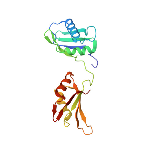
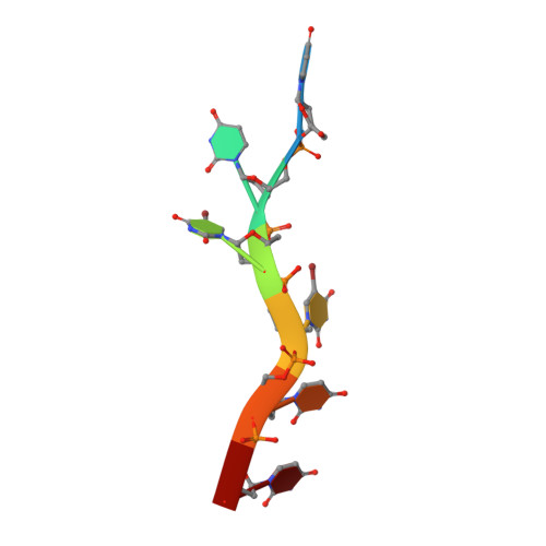
U2AF65 adapts to diverse pre-mRNA splice sites through conformational selection of specific and promiscuous RNA recognition motifs.

Jenkins, J.L.Agrawal, A.A.Gupta, A.Green, M.R.Kielkopf, C.L.

(2013) Nucleic Acids Res 41: 3859-3873

  • DOI: https://doi.org/10.1093/nar/gkt046
  • Primary Citation of Related Structures:  
    3VAF, 3VAG, 3VAH, 3VAI, 3VAJ, 3VAK, 3VAL, 3VAM

  • PubMed Abstract: 

    Degenerate splice site sequences mark the intron boundaries of pre-mRNA transcripts in multicellular eukaryotes. The essential pre-mRNA splicing factor U2AF(65) is faced with the paradoxical tasks of accurately targeting polypyrimidine (Py) tracts preceding 3' splice sites while adapting to both cytidine and uridine nucleotides with nearly equivalent frequencies. To understand how U2AF(65) recognizes degenerate Py tracts, we determined six crystal structures of human U2AF(65) bound to cytidine-containing Py tracts. As deoxy-ribose backbones were required for co-crystallization with these Py tracts, we also determined two baseline structures of U2AF(65) bound to the deoxy-uridine counterparts and compared the original, RNA-bound structure. Local structural changes suggest that the N-terminal RNA recognition motif 1 (RRM1) is more promiscuous for cytosine-containing Py tracts than the C-terminal RRM2. These structural differences between the RRMs were reinforced by the specificities of wild-type and site-directed mutant U2AF(65) for region-dependent cytosine- and uracil-containing RNA sites. Small-angle X-ray scattering analyses further demonstrated that Py tract variations select distinct inter-RRM spacings from a pre-existing ensemble of U2AF(65) conformations. Our results highlight both local and global conformational selection as a means for universal 3' splice site recognition by U2AF(65).


  • Organizational Affiliation

    Center for RNA Biology and Department of Biochemistry and Biophysics, University of Rochester School of Medicine and Dentistry, Rochester, NY 14642, USA.


Macromolecules

Find similar proteins by:  (by identity cutoff)  |  3D Structure
Entity ID: 1
MoleculeChains Sequence LengthOrganismDetailsImage
Splicing factor U2AF 65 kDa subunit
A, B
174Homo sapiensMutation(s): 0 
Gene Names: U2AF2U2AF65
UniProt & NIH Common Fund Data Resources
Find proteins for P26368 (Homo sapiens)
Explore P26368 
Go to UniProtKB:  P26368
PHAROS:  P26368
GTEx:  ENSG00000063244 
Entity Groups  
Sequence Clusters30% Identity50% Identity70% Identity90% Identity95% Identity100% Identity
UniProt GroupP26368
Sequence Annotations
Expand
  • Reference Sequence

Find similar nucleic acids by:  Sequence   |   3D Structure  

Entity ID: 2
MoleculeChains LengthOrganismImage
DNA 5'-D(*UP*UP*(BRU)P*(BRU)P*UP*UP*U)-3'C [auth P],
D [auth E]
7N/A
Sequence Annotations
Expand
  • Reference Sequence
Small Molecules
Ligands 4 Unique
IDChains Name / Formula / InChI Key2D Diagram3D Interactions
CPQ
Query on CPQ

Download Ideal Coordinates CCD File 
J [auth B]N,N-BIS(3-D-GLUCONAMIDOPROPYL)DEOXYCHOLAMIDE
C42 H75 N3 O15
OJSUWTDDXLCUFR-HGZMBBKESA-N
SO4
Query on SO4

Download Ideal Coordinates CCD File 
G [auth A]
H [auth A]
M [auth B]
N [auth B]
O [auth B]
G [auth A],
H [auth A],
M [auth B],
N [auth B],
O [auth B],
P
SULFATE ION
O4 S
QAOWNCQODCNURD-UHFFFAOYSA-L
GOL
Query on GOL

Download Ideal Coordinates CCD File 
I [auth A]GLYCEROL
C3 H8 O3
PEDCQBHIVMGVHV-UHFFFAOYSA-N
DIO
Query on DIO

Download Ideal Coordinates CCD File 
E [auth A],
F [auth A],
K [auth B],
L [auth B]
1,4-DIETHYLENE DIOXIDE
C4 H8 O2
RYHBNJHYFVUHQT-UHFFFAOYSA-N
Experimental Data & Validation

Experimental Data

  • Method: X-RAY DIFFRACTION
  • Resolution: 2.49 Å
  • R-Value Free: 0.289 
  • R-Value Work: 0.231 
  • R-Value Observed: 0.236 
  • Space Group: C 1 2 1
Unit Cell:
Length ( Å )Angle ( ˚ )
a = 164.296α = 90
b = 36.981β = 125.67
c = 99.431γ = 90
Software Package:
Software NamePurpose
DENZOdata reduction
SCALEPACKdata scaling
REFMACrefinement
PDB_EXTRACTdata extraction
HKL-2000data collection
PHASERphasing

Structure Validation

View Full Validation Report


Ligand Structure Quality Assessment 


Entry History 

Deposition Data

Revision History  (Full details and data files)

  • Version 1.0: 2013-02-13
    Type: Initial release
  • Version 1.1: 2013-05-22
    Changes: Database references
  • Version 1.2: 2017-08-16
    Changes: Refinement description, Source and taxonomy
  • Version 1.3: 2023-09-13
    Changes: Data collection, Database references, Derived calculations, Refinement description