3V6M | pdb_00003v6m

Inhibition of caspase-6 activity by single mutation outside the active site


Experimental Data Snapshot

  • Method: X-RAY DIFFRACTION
  • Resolution: 2.69 Å
  • R-Value Free: 
    0.259 (Depositor), 0.253 (DCC) 
  • R-Value Work: 
    0.215 (Depositor), 0.211 (DCC) 
  • R-Value Observed: 
    0.217 (Depositor) 

Starting Model: experimental
View more details

wwPDB Validation 3D Report Full Report

Validation slider image for 3V6M

This is version 1.2 of the entry. See complete history

Literature

Inhibitory mechanism of caspase-6 phosphorylation revealed by crystal structures, molecular dynamics simulations, and biochemical assays

Cao, Q.Wang, X.J.Liu, C.W.Liu, D.F.Li, L.F.Gao, Y.Q.Su, X.D.

(2012) J Biological Chem 287: 15371-15379

  • DOI: https://doi.org/10.1074/jbc.M112.351213
  • Primary Citation Related Structures: 
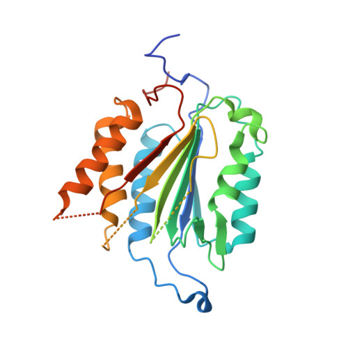
    3V6L, 3V6M

  • PubMed Abstract: 

    The apoptotic effector caspase-6 (CASP6) has been clearly identified as a drug target due to its strong association with neurodegeneration and axonal pruning events as well as its crucial roles in Huntington disease and Alzheimer disease. CASP6 activity is suppressed by ARK5-mediated phosphorylation at Ser(257) with an unclear mechanism. In this work, we solved crystal structures of ΔproCASP6S257E and p20/p10S257E, which mimicked the phosphorylated CASP6 zymogen and activated CASP6, respectively. The structural investigation combined with extensive biochemical assay and molecular dynamics simulation studies revealed that phosphorylation on Ser(257) inhibited self-activation of CASP6 zymogen by "locking" the enzyme in the TEVD(193)-bound "inhibited state." The structural and biochemical results also showed that phosphorylation on Ser(257) inhibited the CASP6 activity by steric hindrance. These results disclosed the inhibition mechanism of CASP6 phosphorylation and laid the foundation for a new strategy of rational CASP6 drug design.


  • Organizational Affiliation
    • State Key Laboratory of Protein and Plant Gene Research and Biodynamic Optical Imaging Center (BIOPIC), School of Life Sciences, Peking University, Beijing 100871, China.

Macromolecule Content 

  • Total Structure Weight: 257.82 kDa 
  • Atom Count: 12,783 
  • Modeled Residue Count: 1,635 
  • Deposited Residue Count: 2,232 
  • Unique protein chains: 1

Macromolecules

Find similar proteins by:|  3D Structure
Entity ID: 1
MoleculeChains  Sequence LengthOrganismDetailsImage
Caspase-6279Homo sapiensMutation(s): 0 
Gene Names: CASP6
EC: 3.4.22.59
UniProt & NIH Common Fund Data Resources
Find proteins for P55212 (Homo sapiens)
Explore P55212 
Go to UniProtKB:  P55212
PHAROS:  P55212
GTEx:  ENSG00000138794 
Entity Groups
Sequence Clusters30% Identity50% Identity70% Identity90% Identity95% Identity100% Identity
UniProt GroupP55212
Sequence Annotations
Expand
Reference Sequence

Experimental Data & Validation

Experimental Data

  • Method: X-RAY DIFFRACTION
  • Resolution: 2.69 Å
  • R-Value Free:  0.259 (Depositor), 0.253 (DCC) 
  • R-Value Work:  0.215 (Depositor), 0.211 (DCC) 
  • R-Value Observed: 0.217 (Depositor) 
Space Group: P 1 21 1
Unit Cell:
Length ( Å )Angle ( ˚ )
a = 81.146α = 90
b = 162.63β = 95.01
c = 89.15γ = 90
Software Package:
Software NamePurpose
HKL-2000data collection
PHENIXmodel building
PHENIXrefinement
HKL-2000data reduction
HKL-2000data scaling
PHENIXphasing

Structure Validation

View Full Validation Report


Entry History 

Deposition Data

Revision History  (Full details and data files)

  • Version 1.0: 2012-03-28
    Type: Initial release
  • Version 1.1: 2012-06-13
    Changes: Database references, Structure summary
  • Version 1.2: 2023-11-08
    Changes: Data collection, Database references, Refinement description