3V5X

Structure of FBXL5 hemerythrin domain, C2 cell


Experimental Data Snapshot

  • Method: X-RAY DIFFRACTION
  • Resolution: 1.85 Å
  • R-Value Free: 0.209 
  • R-Value Work: 0.169 
  • R-Value Observed: 0.171 

wwPDB Validation   3D Report Full Report


This is version 1.4 of the entry. See complete history


Literature

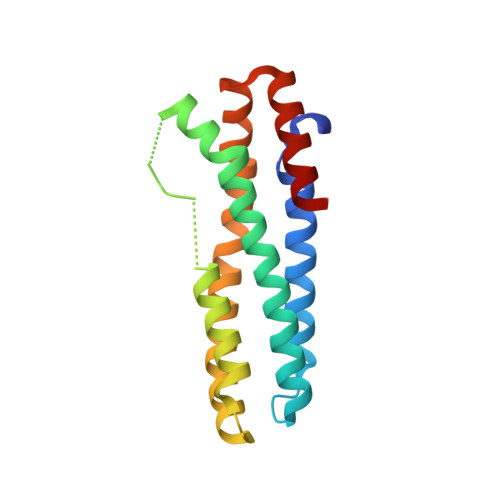
Structural and Molecular Characterization of Iron-sensing Hemerythrin-like Domain within F-box and Leucine-rich Repeat Protein 5 (FBXL5).

Thompson, J.W.Salahudeen, A.A.Chollangi, S.Ruiz, J.C.Brautigam, C.A.Makris, T.M.Lipscomb, J.D.Tomchick, D.R.Bruick, R.K.

(2012) J Biol Chem 287: 7357-7365

  • DOI: https://doi.org/10.1074/jbc.M111.308684
  • Primary Citation of Related Structures:  
    3V5X, 3V5Y, 3V5Z

  • PubMed Abstract: 

    Mammalian cells maintain iron homeostasis by sensing changes in bioavailable iron levels and promoting adaptive responses. FBXL5 is a subunit of an E3 ubiquitin ligase complex that mediates the stability of iron regulatory protein 2, an important posttranscriptional regulator of several genes involved in iron metabolism. The stability of FBXL5 is regulated in an iron- and oxygen-responsive manner, contingent upon the presence of its N-terminal domain. Here we present the atomic structure of the FBXL5 N terminus, a hemerythrin-like α-helical bundle fold not previously observed in mammalian proteins. The core of this domain employs an unusual assortment of amino acids necessary for the assembly and sensing properties of its diiron center. These regulatory features govern the accessibility of a mapped sequence required for proteasomal degradation of FBXL5. Detailed molecular and structural characterization of the ligand-responsive hemerythrin domain provides insights into the mechanisms by which FBXL5 serves as a unique mammalian metabolic sensor.


  • Organizational Affiliation

    Department of Biochemistry, University of Texas Southwestern Medical Center at Dallas, Dallas, Texas 75390, USA.


Macromolecules
Find similar proteins by:  (by identity cutoff)  |  3D Structure
Entity ID: 1
MoleculeChains Sequence LengthOrganismDetailsImage
F-box/LRR-repeat protein 5
A, B
161Homo sapiensMutation(s): 0 
Gene Names: FBL4FBL5FBXL5FLR1
UniProt & NIH Common Fund Data Resources
Find proteins for Q9UKA1 (Homo sapiens)
Explore Q9UKA1 
Go to UniProtKB:  Q9UKA1
PHAROS:  Q9UKA1
GTEx:  ENSG00000118564 
Entity Groups  
Sequence Clusters30% Identity50% Identity70% Identity90% Identity95% Identity100% Identity
UniProt GroupQ9UKA1
Sequence Annotations
Expand
  • Reference Sequence
Experimental Data & Validation

Experimental Data

  • Method: X-RAY DIFFRACTION
  • Resolution: 1.85 Å
  • R-Value Free: 0.209 
  • R-Value Work: 0.169 
  • R-Value Observed: 0.171 
  • Space Group: C 1 2 1
Unit Cell:
Length ( Å )Angle ( ˚ )
a = 60.118α = 90
b = 75.802β = 111.75
c = 78.727γ = 90
Software Package:
Software NamePurpose
DENZOdata reduction
SCALEPACKdata scaling
PHENIXrefinement
PDB_EXTRACTdata extraction
SBC-Collectdata collection
HKL-3000data reduction
HKL-3000data scaling
SHELXDphasing
MLPHAREphasing

Structure Validation

View Full Validation Report


Entry History 

Deposition Data

Revision History  (Full details and data files)

  • Version 1.0: 2012-01-25
    Type: Initial release
  • Version 1.1: 2012-02-01
    Changes: Database references
  • Version 1.2: 2012-03-21
    Changes: Database references
  • Version 1.3: 2017-11-08
    Changes: Refinement description
  • Version 1.4: 2024-02-28
    Changes: Data collection, Database references, Derived calculations