3V4X

The Biochemical and Structural Basis for Inhibition of Enterococcus faecalis HMG-CoA Synthase, mvaS, by Hymeglusin


Experimental Data Snapshot

  • Method: X-RAY DIFFRACTION
  • Resolution: 1.95 Å
  • R-Value Free: 0.218 
  • R-Value Work: 0.171 
  • R-Value Observed: 0.175 

wwPDB Validation   3D Report Full Report


Ligand Structure Quality Assessment 


This is version 1.2 of the entry. See complete history


Literature

Biochemical and structural basis for inhibition of Enterococcus faecalis hydroxymethylglutaryl-CoA synthase, mvaS, by hymeglusin.

Skaff, D.A.Ramyar, K.X.McWhorter, W.J.Barta, M.L.Geisbrecht, B.V.Miziorko, H.M.

(2012) Biochemistry 51: 4713-4722

  • DOI: https://doi.org/10.1021/bi300037k
  • Primary Citation of Related Structures:  
    3V4N, 3V4X

  • PubMed Abstract: 

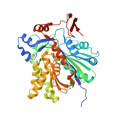
    Hymeglusin (1233A, F244, L-659-699) is established as a specific β-lactone inhibitor of eukaryotic hydroxymethylglutaryl-CoA synthase (HMGCS). Inhibition results from formation of a thioester adduct to the active site cysteine. In contrast, the effects of hymeglusin on bacterial HMG-CoA synthase, mvaS, have been minimally characterized. Hymeglusin blocks growth of Enterococcus faecalis. After removal of the inhibitor from culture media, a growth curve inflection point at 3.1 h is observed (vs 0.7 h for the uninhibited control). Upon hymeglusin inactivation of purified E. faecalis mvaS, the thioester adduct is more stable than that measured for human HMGCS. Hydroxylamine cleaves the thioester adduct; substantial enzyme activity is restored at a rate that is 8-fold faster for human HMGCS than for mvaS. Structural results explain these differences in enzyme-inhibitor thioester adduct stability and solvent accessibility. The E. faecalis mvaS-hymeglusin cocrystal structure (1.95 Å) reveals virtually complete occlusion of the bound inhibitor in a narrow tunnel that is largely sequestered from bulk solvent. In contrast, eukaryotic (Brassica juncea) HMGCS binds hymeglusin in a more solvent-exposed cavity.


  • Organizational Affiliation

    University of Missouri, Kansas City, MO, USA.


Macromolecules
Find similar proteins by:  (by identity cutoff)  |  3D Structure
Entity ID: 1
MoleculeChains Sequence LengthOrganismDetailsImage
HMG-CoA synthase
A, B, C, D
388Enterococcus faecalisMutation(s): 1 
Gene Names: mvaS
EC: 2.3.3.10
UniProt
Find proteins for Q9FD71 (Enterococcus faecalis)
Explore Q9FD71 
Go to UniProtKB:  Q9FD71
Entity Groups  
Sequence Clusters30% Identity50% Identity70% Identity90% Identity95% Identity100% Identity
UniProt GroupQ9FD71
Sequence Annotations
Expand
  • Reference Sequence
Small Molecules
Binding Affinity Annotations 
IDSourceBinding Affinity
F24 PDBBind:  3V4X Ki: 700 (nM) from 1 assay(s)
Experimental Data & Validation

Experimental Data

  • Method: X-RAY DIFFRACTION
  • Resolution: 1.95 Å
  • R-Value Free: 0.218 
  • R-Value Work: 0.171 
  • R-Value Observed: 0.175 
  • Space Group: P 1 21 1
Unit Cell:
Length ( Å )Angle ( ˚ )
a = 108.333α = 90
b = 52.756β = 99.51
c = 117.576γ = 90
Software Package:
Software NamePurpose
HKL-2000data collection
PHENIXmodel building
PHENIXrefinement
HKL-2000data reduction
HKL-2000data scaling
PHENIXphasing

Structure Validation

View Full Validation Report


Ligand Structure Quality Assessment 


Entry History 

Deposition Data

Revision History  (Full details and data files)

  • Version 1.0: 2012-04-25
    Type: Initial release
  • Version 1.1: 2013-11-27
    Changes: Database references
  • Version 1.2: 2023-09-13
    Changes: Data collection, Database references, Derived calculations, Refinement description