3V4R | pdb_00003v4r

Crystal structure of a UvrB dimer-DNA complex


Experimental Data Snapshot

  • Method: X-RAY DIFFRACTION
  • Resolution: 3.25 Å
  • R-Value Free: 
    0.219 (Depositor), 0.238 (DCC) 
  • R-Value Work: 
    0.180 (Depositor), 0.193 (DCC) 
  • R-Value Observed: 
    0.182 (Depositor) 

Starting Model: experimental
View more details

wwPDB Validation 3D Report Full Report

Validation slider image for 3V4R

Ligand Structure Quality Assessment 


This is version 1.2 of the entry. See complete history

Literature

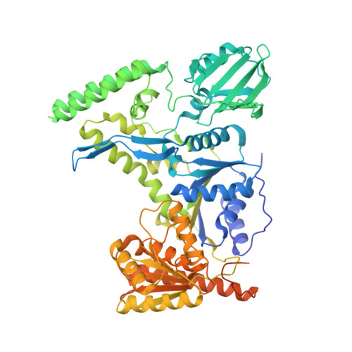
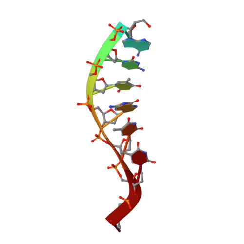
Crystal structure of the UvrB dimer: insights into the nature and functioning of the UvrAB damage engagement and UvrB-DNA complexes.

Webster, M.P.Jukes, R.Zamfir, V.S.Kay, C.W.Bagneris, C.Barrett, T.

(2012) Nucleic Acids Res 40: 8743-8758

  • DOI: https://doi.org/10.1093/nar/gks633
  • Primary Citation Related Structures: 
    3V4R

  • PubMed Abstract: 

    UvrB has a central role in the highly conserved UvrABC pathway functioning not only as a damage recognition element but also as an essential component of the lesion tracking machinery. While it has been recently confirmed that the tracking assembly comprises a UvrA2B2 heterotetramer, the configurations of the damage engagement and UvrB-DNA handover complexes remain obscure. Here, we present the first crystal structure of a UvrB dimer whose biological significance has been verified using both chemical cross-linking and electron paramagnetic resonance spectroscopy. We demonstrate that this dimeric species stably associates with UvrA and forms a UvrA2B2-DNA complex. Our studies also illustrate how signals are transduced between the ATP and DNA binding sites to generate the helicase activity pivotal to handover and formation of the UvrB2-DNA complex, providing key insights into the configurations of these important repair intermediates.


  • Organizational Affiliation
    • Department of Biological Sciences, Institute of Structural and Molecular Biology, Crystallography, Birkbeck College, Malet Street, London WC1E 7HX, UK.

Macromolecule Content 

  • Total Structure Weight: 160.21 kDa 
  • Atom Count: 8,815 
  • Modeled Residue Count: 1,179 
  • Deposited Residue Count: 1,350 
  • Unique protein chains: 1
  • Unique nucleic acid chains: 1

Macromolecules


Find similar proteins by:|  3D Structure
Entity ID: 1
MoleculeChains  Sequence LengthOrganismDetailsImage
UvrABC system protein B
A, B
667Bacillus subtilisMutation(s): 0 
Gene Names: BSU35170dinAuvruvrB
EC: 3.1.21.5
UniProt
Find proteins for P37954 (Bacillus subtilis (strain 168))
Explore P37954 
Go to UniProtKB:  P37954
Entity Groups
Sequence Clusters30% Identity50% Identity70% Identity90% Identity95% Identity100% Identity
UniProt GroupP37954
Sequence Annotations
Expand
Reference Sequence
Find similar nucleic acids by:  Sequence
Entity ID: 2
MoleculeChains LengthOrganismImage
DNA: 5 -TACTGTTT-3
C, D
8N/A
Sequence Annotations
Expand
Reference Sequence

Experimental Data & Validation

Experimental Data

  • Method: X-RAY DIFFRACTION
  • Resolution: 3.25 Å
  • R-Value Free:  0.219 (Depositor), 0.238 (DCC) 
  • R-Value Work:  0.180 (Depositor), 0.193 (DCC) 
  • R-Value Observed: 0.182 (Depositor) 
Space Group: P 21 21 21
Unit Cell:
Length ( Å )Angle ( ˚ )
a = 95.94α = 90
b = 100.31β = 90
c = 163.63γ = 90
Software Package:
Software NamePurpose
DNAdata collection
PHASERphasing
BUSTERrefinement
XDSdata reduction
XSCALEdata scaling

Structure Validation

View Full Validation Report


Ligand Structure Quality Assessment 


Entry History 

Deposition Data

Revision History  (Full details and data files)

  • Version 1.0: 2012-07-04
    Type: Initial release
  • Version 1.1: 2012-10-10
    Changes: Database references
  • Version 1.2: 2023-09-13
    Changes: Data collection, Database references, Derived calculations, Refinement description