3V4A | pdb_00003v4a

Structure of ar lbd with activator peptide and sarm inhibitor 2


Experimental Data Snapshot

  • Method: X-RAY DIFFRACTION
  • Resolution: 1.95 Å
  • R-Value Work: 
    0.172 (Depositor), 0.169 (DCC) 
  • R-Value Observed: 
    0.172 (Depositor) 

Starting Model: experimental
View more details

wwPDB Validation 3D Report Full Report

Validation slider image for 3V4A

Ligand Structure Quality Assessment 


This is version 1.4 of the entry. See complete history

Literature

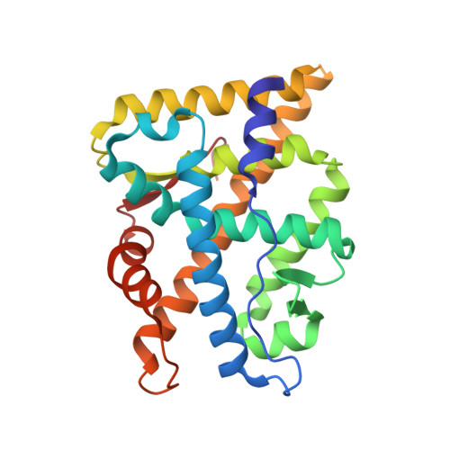
Discovery of diarylhydantoins as new selective androgen receptor modulators.

Nique, F.Hebbe, S.Peixoto, C.Annoot, D.Lefrancois, J.-M.Duval, E.Michoux, L.Triballeau, N.Lemoullec, J.M.Mollat, P.Thauvin, M.Prange, T.Minet, D.Clement-Lacroix, P.Robin-Jagerschmidt, C.Fleury, D.Guedin, D.Deprez, P.

(2012) J Med Chem 55: 8225-8235

  • DOI: https://doi.org/10.1021/jm300249m
  • Primary Citation Related Structures: 
    3V49, 3V4A

  • PubMed Abstract: 

    A novel selective androgen receptor modulator scaffold has been discovered through structural modifications of hydantoin antiandrogens. Several 4-(4-hydroxyphenyl)-N-arylhydantoins displayed partial agonism with nanomolar in vitro potency in transactivation experiments using androgen receptor (AR) transfected cells. In a standard castrated male rat model, several compounds showed good anabolic activity on levator ani muscle, dissociated from the androgenic activity on ventral prostate, after oral dosing at 30 mg/kg. (+)-4-[3,4-Dimethyl-2,5-dioxo-4-(4-hydroxyphenyl)imidazolidin-1-yl]-2-(trifluoromethyl)benzonitrile ((+)-11b) displayed anabolic potency with a strong dissociation between levator ani muscle and ventral prostate (A(50) = 0.5 mg/kg vs 70 mg/kg). The binding modes of two compounds, including (+)-11b, within the AR ligand-binding domain have been studied by cocrystallization experiments using a coactivator-like peptide. Both compounds bound to the same site, and the overall structures of the AR were very similar.


  • Organizational Affiliation
    • GALAPAGOS, Parc Biocitech, 102 Avenue Gaston Roussel, 93230 Romainville, France.

Macromolecule Content 

  • Total Structure Weight: 30.79 kDa 
  • Atom Count: 2,374 
  • Modeled Residue Count: 255 
  • Deposited Residue Count: 260 
  • Unique protein chains: 2

Macromolecules

Find similar proteins by:|  3D Structure
Entity ID: 1
MoleculeChains  Sequence LengthOrganismDetailsImage
Androgen receptor249Homo sapiensMutation(s): 0 
Gene Names: ARDHTRNR3C4
UniProt & NIH Common Fund Data Resources
Find proteins for P10275 (Homo sapiens)
Explore P10275 
Go to UniProtKB:  P10275
PHAROS:  P10275
GTEx:  ENSG00000169083 
Entity Groups
Sequence Clusters30% Identity50% Identity70% Identity90% Identity95% Identity100% Identity
UniProt GroupP10275
Sequence Annotations
Expand
Reference Sequence
Find similar proteins by:  Sequence   |   3D Structure  
Entity ID: 2
MoleculeChains  Sequence LengthOrganismDetailsImage
Androgen receptor11Homo sapiensMutation(s): 0 
UniProt & NIH Common Fund Data Resources
Find proteins for P10275 (Homo sapiens)
Explore P10275 
Go to UniProtKB:  P10275
PHAROS:  P10275
GTEx:  ENSG00000169083 
Entity Groups
Sequence Clusters30% Identity50% Identity70% Identity90% Identity95% Identity100% Identity
UniProt GroupP10275
Sequence Annotations
Expand
Reference Sequence

Small Molecules

Ligands 2 Unique
IDChains Name / Formula / InChI Key2D Diagram3D Interactions
PK1

Query on PK1


Download:Ideal Coordinates CCD File
C [auth A](5R)-3-(3,4-dichlorophenyl)-5-(4-hydroxyphenyl)-1,5-dimethyl-2-thioxoimidazolidin-4-one
C17 H14 Cl2 N2 O2 S
YPONBLYNQFHCLA-QGZVFWFLSA-N
SO4

Query on SO4


Download:Ideal Coordinates CCD File
D [auth A]SULFATE ION
O4 S
QAOWNCQODCNURD-UHFFFAOYSA-L
Binding Affinity Annotations 
IDSourceBinding Affinity
PK1 BindingDB:  3V4A EC50: 0.8 (nM) from 1 assay(s)

Experimental Data & Validation

Experimental Data

  • Method: X-RAY DIFFRACTION
  • Resolution: 1.95 Å
  • R-Value Work:  0.172 (Depositor), 0.169 (DCC) 
  • R-Value Observed: 0.172 (Depositor) 
Space Group: P 21 21 21
Unit Cell:
Length ( Å )Angle ( ˚ )
a = 54.446α = 90
b = 66.805β = 90
c = 69.854γ = 90
Software Package:
Software NamePurpose
ADSCdata collection
SHELXmodel building
SHELXL-97refinement
MOSFLMdata reduction
SCALAdata scaling
SHELXphasing

Structure Validation

View Full Validation Report


Ligand Structure Quality Assessment 


Entry History 

Revision History  (Full details and data files)

  • Version 1.0: 2012-09-05
    Type: Initial release
  • Version 1.1: 2012-12-19
    Changes: Database references
  • Version 1.2: 2014-10-08
    Changes: Structure summary
  • Version 1.3: 2018-01-24
    Changes: Structure summary
  • Version 1.4: 2023-09-13
    Changes: Data collection, Database references, Derived calculations, Refinement description