3V2A | pdb_00003v2a

VEGFR-2/VEGF-A COMPLEX STRUCTURE


Experimental Data Snapshot

  • Method: X-RAY DIFFRACTION
  • Resolution: 3.20 Å
  • R-Value Free: 
    0.300 (Depositor), 0.307 (DCC) 
  • R-Value Work: 
    0.244 (Depositor), 0.261 (DCC) 
  • R-Value Observed: 
    0.247 (Depositor) 

Starting Model: experimental
View more details

wwPDB Validation 3D Report Full Report

Validation slider image for 3V2A

This is version 1.4 of the entry. See complete history

Literature

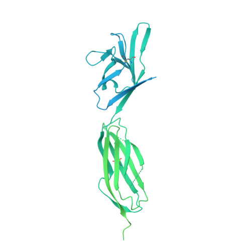
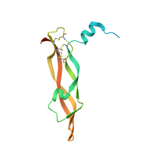
Thermodynamic and structural description of allosterically regulated VEGFR-2 dimerization.

Brozzo, M.S.Bjelic, S.Kisko, K.Schleier, T.Leppanen, V.M.Alitalo, K.Winkler, F.K.Ballmer-Hofer, K.

(2012) Blood 119: 1781-1788

  • DOI: https://doi.org/10.1182/blood-2011-11-390922
  • Primary Citation Related Structures: 
    3V2A, 3V6B

  • PubMed Abstract: 

    VEGFs activate 3 receptor tyrosine kinases, VEGFR-1, VEGFR-2, and VEGFR-3, promoting angiogenic and lymphangiogenic signaling. The extracellular receptor domain (ECD) consists of 7 Ig-homology domains; domains 2 and 3 (D23) represent the ligand-binding domain, whereas the function of D4-7 is unclear. Ligand binding promotes receptor dimerization and instigates transmembrane signaling and receptor kinase activation. In the present study, isothermal titration calorimetry showed that the Gibbs free energy of VEGF-A, VEGF-C, or VEGF-E binding to D23 or the full-length ECD of VEGFR-2 is dominated by favorable entropic contribution with enthalpic penalty. The free energy of VEGF binding to the ECD is 1.0-1.7 kcal/mol less favorable than for binding to D23. A model of the VEGF-E/VEGFR-2 ECD complex derived from small-angle scattering data provided evidence for homotypic interactions in D4-7. We also solved the crystal structures of complexes between VEGF-A or VEGF-E with D23, which revealed comparable binding surfaces and similar interactions between the ligands and the receptor, but showed variation in D23 twist angles. The energetically unfavorable homotypic interactions in D4-7 may be required for re-orientation of receptor monomers, and this mechanism might prevent ligand-independent activation of VEGFR-2 to evade the deleterious consequences for blood and lymph vessel homeostasis arising from inappropriate receptor activation.


  • Organizational Affiliation
    • Biomolecular Research, Molecular Cell Biology Paul Scherrer Institut, Villigen, Switzerland.

Macromolecule Content 

  • Total Structure Weight: 102.11 kDa 
  • Atom Count: 2,114 
  • Modeled Residue Count: 280 
  • Deposited Residue Count: 906 
  • Unique protein chains: 2

Macromolecules

Find similar proteins by:|  3D Structure
Entity ID: 1
MoleculeChains  Sequence LengthOrganismDetailsImage
Vascular endothelial growth factor receptor 2A [auth R]772Homo sapiensMutation(s): 0 
Gene Names: FLK1KDRVEGFR2
EC: 2.7.10.1
UniProt & NIH Common Fund Data Resources
Find proteins for P35968 (Homo sapiens)
Explore P35968 
Go to UniProtKB:  P35968
PHAROS:  P35968
GTEx:  ENSG00000128052 
Entity Groups
Sequence Clusters30% Identity50% Identity70% Identity90% Identity95% Identity100% Identity
UniProt GroupP35968
Sequence Annotations
Expand
Reference Sequence
Find similar proteins by:|  3D Structure
Entity ID: 2
MoleculeChains  Sequence LengthOrganismDetailsImage
Vascular endothelial growth factor AB [auth A]134Homo sapiensMutation(s): 0 
Gene Names: VEGFVEGF-AVEGFA
UniProt & NIH Common Fund Data Resources
Find proteins for P15692 (Homo sapiens)
Explore P15692 
Go to UniProtKB:  P15692
PHAROS:  P15692
GTEx:  ENSG00000112715 
Entity Groups
Sequence Clusters30% Identity50% Identity70% Identity90% Identity95% Identity100% Identity
UniProt GroupP15692
Sequence Annotations
Expand
Reference Sequence

Experimental Data & Validation

Experimental Data

  • Method: X-RAY DIFFRACTION
  • Resolution: 3.20 Å
  • R-Value Free:  0.300 (Depositor), 0.307 (DCC) 
  • R-Value Work:  0.244 (Depositor), 0.261 (DCC) 
  • R-Value Observed: 0.247 (Depositor) 
Space Group: P 65 2 2
Unit Cell:
Length ( Å )Angle ( ˚ )
a = 80.969α = 90
b = 80.969β = 90
c = 332.063γ = 120
Software Package:
Software NamePurpose
RemDAqdata collection
PHASERphasing
PHENIXrefinement
XDSdata reduction
XSCALEdata scaling

Structure Validation

View Full Validation Report


Entry History 

Deposition Data

Revision History  (Full details and data files)

  • Version 1.0: 2012-01-18
    Type: Initial release
  • Version 1.1: 2012-03-07
    Changes: Database references
  • Version 1.2: 2017-11-08
    Changes: Refinement description
  • Version 1.3: 2024-04-03
    Changes: Data collection, Database references, Refinement description
  • Version 1.4: 2024-11-20
    Changes: Structure summary