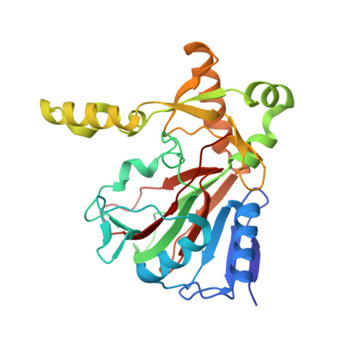
The Fe(II) / alpha-ketoglutarate dependent taurine dioxygenases from Pseudomonas putida and Escherichia coli are tetramers
Knauer, S.H., Hartl-Spiegelhauer, O., Schwarzinger, S., Hanzelmann, P., Dobbek, H.(2012) FEBS J 279: 816-831
- PubMed: 22221834 Search on PubMed
- DOI: https://doi.org/10.1111/j.1742-4658.2012.08473.x
- Primary Citation Related Structures:
3V15, 3V17 - PubMed Abstract:
Fe(II)/α-ketoglutarate-dependent oxygenases are versatile catalysts associated with a number of different biological functions in which they use the oxidizing power of activated dioxygen to convert a variety of substrates. A mononuclear nonheme iron center is used to couple the decarboxylation of the cosubstrate α-ketoglutarate with a two-electron oxidation of the substrate, which is a hydroxylation in most cases. Although Fe(II)/α-ketoglutarate-dependent oxygenases have diverse amino acid sequences and substrate specifity, it is assumed that they share a common mechanism. One representative of this enzyme family is the Fe(II)/α-ketoglutarate-dependent taurine dioxygenase that catalyzes the hydroxylation of taurine yielding sulfite and aminoacetaldehyde. Its mechanism has been studied in detail becoming a model system for the whole enzyme family. However, its oligomeric state and architecture have been disputed. Here, we report the biochemical and kinetic characterization of the Fe(II)/α-ketoglutarate-dependent taurine dioxygenase from Pseudomonas putida KT2440 (TauD(Pp) ). We also present three crystal structures of the apo form of this enzyme. Comparisons with taurine dioxygenase from Escherichia coli (TauD(Ec) ) demonstrate that both enzymes are quite similar regarding their spectra, structure and kinetics, and only minor differences for the accumulation of intermediates during the reaction have been observed. Structural data and analytical gel filtration, as well as sedimentation velocity analytical ultracentrifugation, show that both TauD(Pp) and TauD(Ec) are tetramers in solution and in the crystals, which is in contrast to the earlier description of taurine dioxygenase from E. coli as a dimer. Database The atomic coordinates and structure factors have been deposited with the Brookhaven Protein Data Bank (entry 3PVJ, 3V15, 3V17) Structured digital abstract • tauDpp and tauDpp bind by molecular sieving (View interaction) • tauDpp and tauDpp bind by x-ray crystallography (View interaction) • tauDEc and tauDEc bind by molecular sieving (View interaction).
- Institut für Biologie, Strukturbiologie/Biochemie, Humboldt-Universität zu Berlin, Germany.
Organizational Affiliation: