3V0H

Crystal structure of Ciona intestinalis voltage sensor-containing phosphatase (Ci-VSP), residues 241-576(C363S), complexed with D-MYO-inositol-1,4,5-triphosphate


Experimental Data Snapshot

  • Method: X-RAY DIFFRACTION
  • Resolution: 1.85 Å
  • R-Value Free: 0.222 
  • R-Value Work: 0.181 
  • R-Value Observed: 0.183 

wwPDB Validation   3D Report Full Report


This is version 1.3 of the entry. See complete history


Literature

A glutamate switch controls voltage-sensitive phosphatase function.

Liu, L.Kohout, S.C.Xu, Q.Muller, S.Kimberlin, C.R.Isacoff, E.Y.Minor, D.L.

(2012) Nat Struct Mol Biol 19: 633-641

  • DOI: https://doi.org/10.1038/nsmb.2289
  • Primary Citation of Related Structures:  
    3V0D, 3V0E, 3V0F, 3V0G, 3V0H, 3V0I, 3V0J

  • PubMed Abstract: 

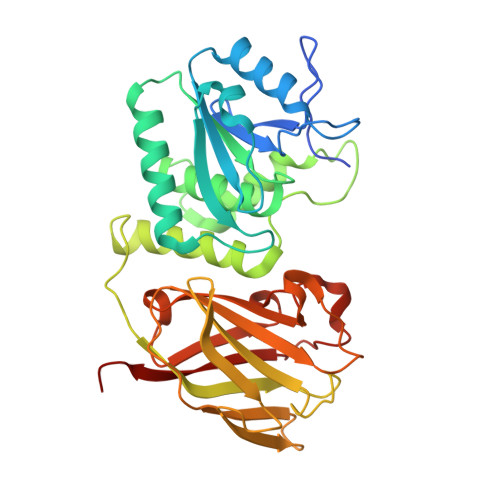
    The Ciona intestinalis voltage-sensing phosphatase (Ci-VSP) couples a voltage-sensing domain (VSD) to a lipid phosphatase that is similar to the tumor suppressor PTEN. How the VSD controls enzyme function has been unclear. Here, we present high-resolution crystal structures of the Ci-VSP enzymatic domain that reveal conformational changes in a crucial loop, termed the 'gating loop', that controls access to the active site by a mechanism in which residue Glu411 directly competes with substrate. Structure-based mutations that restrict gating loop conformation impair catalytic function and demonstrate that Glu411 also contributes to substrate selectivity. Structure-guided mutations further define an interaction between the gating loop and linker that connects the phosphatase to the VSD for voltage control of enzyme activity. Together, the data suggest that functional coupling between the gating loop and the linker forms the heart of the regulatory mechanism that controls voltage-dependent enzyme activation.


  • Organizational Affiliation

    Cardiovascular Research Institute, University of California, San Francisco, San Francisco, California, USA.


Macromolecules
Find similar proteins by:  (by identity cutoff)  |  3D Structure
Entity ID: 1
MoleculeChains Sequence LengthOrganismDetailsImage
Voltage-sensor containing phosphatase
A, B
339Ciona intestinalisMutation(s): 1 
Gene Names: Ci-VSP
UniProt
Find proteins for Q4W8A1 (Ciona intestinalis)
Explore Q4W8A1 
Go to UniProtKB:  Q4W8A1
Entity Groups  
Sequence Clusters30% Identity50% Identity70% Identity90% Identity95% Identity100% Identity
UniProt GroupQ4W8A1
Sequence Annotations
Expand
  • Reference Sequence
Experimental Data & Validation

Experimental Data

  • Method: X-RAY DIFFRACTION
  • Resolution: 1.85 Å
  • R-Value Free: 0.222 
  • R-Value Work: 0.181 
  • R-Value Observed: 0.183 
  • Space Group: P 1 21 1
Unit Cell:
Length ( Å )Angle ( ˚ )
a = 50.665α = 90
b = 79.852β = 92.44
c = 87.927γ = 90
Software Package:
Software NamePurpose
Blu-Icedata collection
MOLREPphasing
REFMACrefinement
HKL-2000data reduction
HKL-2000data scaling

Structure Validation

View Full Validation Report


Entry History 

Deposition Data

Revision History  (Full details and data files)

  • Version 1.0: 2012-05-09
    Type: Initial release
  • Version 1.1: 2012-07-25
    Changes: Database references
  • Version 1.2: 2014-10-08
    Changes: Structure summary
  • Version 1.3: 2023-09-13
    Changes: Data collection, Database references, Derived calculations, Refinement description