3UYV | pdb_00003uyv

Crystal structure of a glycosylated ice-binding protein (LeIBP) from Arctic yeast


Experimental Data Snapshot

  • Method: X-RAY DIFFRACTION
  • Resolution: 2.43 Å
  • R-Value Free: 
    0.286 (Depositor), 0.304 (DCC) 
  • R-Value Work: 
    0.233 (Depositor) 
  • R-Value Observed: 
    0.236 (Depositor) 

Starting Model: experimental
View more details

wwPDB Validation 3D Report Full Report

Validation slider image for 3UYV

This is version 2.2 of the entry. See complete history

Literature

Structural basis for the antifreeze activity of an ice-binding protein from an Arctic yeast.

Lee, J.H.Park, A.K.Do, H.Park, K.S.Moh, S.H.Chi, Y.M.Kim, H.J.

(2012) J Biological Chem 

  • DOI: https://doi.org/10.1074/jbc.M111.331835
  • Primary Citation Related Structures: 
    3UYU, 3UYV

  • PubMed Abstract: 

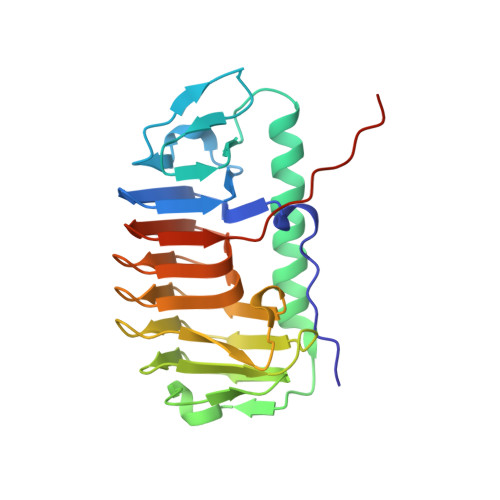
    Arctic yeast Leucosporidium sp. produces a glycosylated ice-binding protein (LeIBP) with a molecular mass of ∼25 kDa, which can lower the freezing point below the melting point once it binds to ice. LeIBP is a member of a large class of ice-binding proteins, the structures of which are unknown. Here, we report the crystal structures of non-glycosylated LeIBP and glycosylated LeIBP at 1.57- and 2.43-Å resolution, respectively. Structural analysis of the LeIBPs revealed a dimeric right-handed β-helix fold, which is composed of three parts: a large coiled structural domain, a long helix region (residues 96-115 form a long α-helix that packs along one face of the β-helix), and a C-terminal hydrophobic loop region ((243)PFVPAPEVV(251)). Unexpectedly, the C-terminal hydrophobic loop region has an extended conformation pointing away from the body of the coiled structural domain and forms intertwined dimer interactions. In addition, structural analysis of glycosylated LeIBP with sugar moieties attached to Asn(185) provides a basis for interpreting previous biochemical analyses as well as the increased stability and secretion of glycosylated LeIBP. We also determined that the aligned Thr/Ser/Ala residues are critical for ice binding within the B face of LeIBP using site-directed mutagenesis. Although LeIBP has a common β-helical fold similar to that of canonical hyperactive antifreeze proteins, the ice-binding site is more complex and does not have a simple ice-binding motif. In conclusion, we could identify the ice-binding site of LeIBP and discuss differences in the ice-binding modes compared with other known antifreeze proteins and ice-binding proteins.


  • Organizational Affiliation
    • Division of Polar Life Sciences, Korea Polar Research Institute, Incheon 406-840, Republic of Korea.

Macromolecule Content 

  • Total Structure Weight: 25.69 kDa 
  • Atom Count: 1,738 
  • Modeled Residue Count: 231 
  • Deposited Residue Count: 241 
  • Unique protein chains: 1

Macromolecules

Find similar proteins by:|  3D Structure
Entity ID: 1
MoleculeChains  Sequence LengthOrganismDetailsImage
Antifreeze protein241Leucosporidium sp. AY30Mutation(s): 0 
Gene Names: AFP
UniProt
Find proteins for C7F6X3 (Leucosporidium sp. (strain AY30))
Explore C7F6X3 
Go to UniProtKB:  C7F6X3
Entity Groups
Sequence Clusters30% Identity50% Identity70% Identity90% Identity95% Identity100% Identity
UniProt GroupC7F6X3
Glycosylation
Glycosylation Sites: 1
Sequence Annotations
Expand
Reference Sequence

Oligosaccharides

Help  
Entity ID: 2
MoleculeChains Length2D Diagram GlycosylationD Interactions
alpha-D-mannopyranose-(1-6)-beta-D-mannopyranose-(1-4)-2-acetamido-2-deoxy-beta-D-glucopyranose-(1-4)-2-acetamido-2-deoxy-beta-D-glucopyranose
B
4N-Glycosylation
Glycosylation Resources
GlyTouCan: G22573RC
GlyCosmos: G22573RC
GlyGen: G22573RC

Experimental Data & Validation

Experimental Data

  • Method: X-RAY DIFFRACTION
  • Resolution: 2.43 Å
  • R-Value Free:  0.286 (Depositor), 0.304 (DCC) 
  • R-Value Work:  0.233 (Depositor) 
  • R-Value Observed: 0.236 (Depositor) 
Space Group: P 65 2 2
Unit Cell:
Length ( Å )Angle ( ˚ )
a = 58.64α = 90
b = 58.64β = 90
c = 292.641γ = 120
Software Package:
Software NamePurpose
PROTEUM PLUSdata collection
MOLREPphasing
REFMACrefinement
MOSFLMdata reduction
SCALAdata scaling

Structure Validation

View Full Validation Report


Entry History 

Deposition Data

Revision History  (Full details and data files)

  • Version 1.0: 2012-02-29
    Type: Initial release
  • Version 1.1: 2020-06-24
    Changes: Advisory, Data collection, Derived calculations
  • Version 2.0: 2020-07-29
    Type: Remediation
    Reason: Carbohydrate remediation
    Changes: Advisory, Atomic model, Data collection, Derived calculations, Structure summary
  • Version 2.1: 2023-11-08
    Changes: Data collection, Database references, Refinement description, Structure summary
  • Version 2.2: 2024-10-30
    Changes: Structure summary