


Crystal structure of DAVA-4
Stetefeld, J.To be published.
Experimental Data Snapshot
Entity ID: 1 | |||||
|---|---|---|---|---|---|
| Molecule | Chains | Sequence Length | Organism | Details | Image |
| Glutamate-1-semialdehyde 2,1-aminomutase | 427 | Synechococcus elongatus PCC 7942 = FACHB-805 | Mutation(s): 0 EC: 5.4.3.8 | 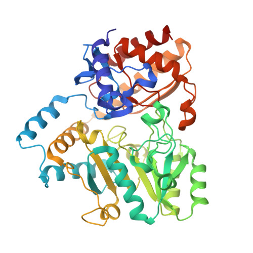 | |
UniProt | |||||
Entity Groups | |||||
| Sequence Clusters | 30% Identity50% Identity70% Identity90% Identity95% Identity100% Identity | ||||
| UniProt Group | Q31QJ2 | ||||
Sequence AnnotationsExpand | |||||
Reference Sequence | |||||
| Ligands 2 Unique | |||||
|---|---|---|---|---|---|
| ID | Chains | Name / Formula / InChI Key | 2D Diagram | 3D Interactions | |
| PLR Download:Ideal Coordinates CCD File | C [auth A], E [auth B] | (5-HYDROXY-4,6-DIMETHYLPYRIDIN-3-YL)METHYL DIHYDROGEN PHOSPHATE C8 H12 N O5 P RBCOYOYDYNXAFA-UHFFFAOYSA-N |  | ||
| HOZ Download:Ideal Coordinates CCD File | D [auth A] | (4S)-4,5-DIAMINOPENTANOIC ACID C5 H12 N2 O2 PQGAAJQIFBEYSA-BYPYZUCNSA-N |  | ||
| Length ( Å ) | Angle ( ˚ ) |
|---|---|
| a = 68.582 | α = 90 |
| b = 108.781 | β = 90 |
| c = 124.712 | γ = 90 |
| Software Name | Purpose |
|---|---|
| CNS | refinement |
| PDB_EXTRACT | data extraction |
| CrystalClear | data collection |
| HKL-2000 | data reduction |
| SCALEPACK | data scaling |
| AMoRE | phasing |


