3UP1 | pdb_00003up1

Crystal structure of the unliganded human interleukin-7 receptor extracellular domain


Experimental Data Snapshot

  • Method: X-RAY DIFFRACTION
  • Resolution: 2.15 Å
  • R-Value Free: 
    0.257 (Depositor), 0.246 (DCC) 
  • R-Value Work: 
    0.214 (Depositor), 0.201 (DCC) 
  • R-Value Observed: 
    0.217 (Depositor) 

wwPDB Validation 3D Report Full Report

Validation slider image for 3UP1

Ligand Structure Quality Assessment 


This is version 2.2 of the entry. See complete history

Literature

Structural reorganization of the interleukin-7 signaling complex.

McElroy, C.A.Holland, P.J.Zhao, P.Lim, J.M.Wells, L.Eisenstein, E.Walsh, S.T.

(2012) Proc Natl Acad Sci U S A 109: 2503-2508

  • DOI: https://doi.org/10.1073/pnas.1116582109
  • Primary Citation Related Structures: 
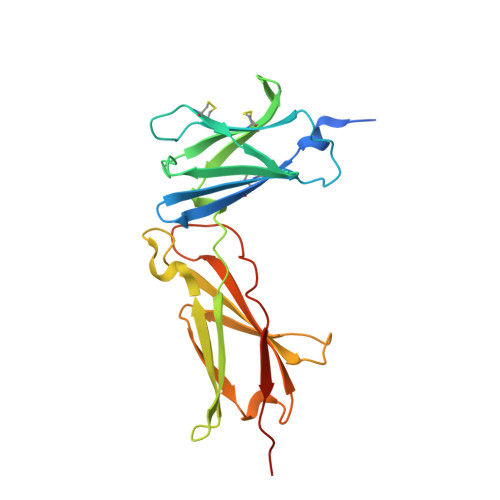
    3UP1

  • PubMed Abstract: 

    We report here an unliganded receptor structure in the common gamma-chain (γ(c)) family of receptors and cytokines. The crystal structure of the unliganded form of the interleukin-7 alpha receptor (IL-7Rα) extracellular domain (ECD) at 2.15 Å resolution reveals a homodimer forming an "X" geometry looking down onto the cell surface with the C termini of the two chains separated by 110 Å and the dimer interface comprising residues critical for IL-7 binding. Further biophysical studies indicate a weak association of the IL-7Rα ECDs but a stronger association between the γ(c)/IL-7Rα ECDs, similar to previous studies of the full-length receptors on CD4(+) T cells. Based on these and previous results, we propose a molecular mechanism detailing the progression from the inactive IL-7Rα homodimer and IL-7Rα-γ(c) heterodimer to the active IL-7-IL-7Rα-γ(c) ternary complex whereby the two receptors undergo at least a 90° rotation away from the cell surface, moving the C termini of IL-7Rα and γ(c) from a distance of 110 Å to less than 30 Å at the cell surface. This molecular mechanism can be used to explain recently discovered IL-7- and γ(c)-independent gain-of-function mutations in IL-7Rα from B- and T-cell acute lymphoblastic leukemia patients. The mechanism may also be applicable to other γ(c) receptors that form inactive homodimers and heterodimers independent of their cytokines.


  • Organizational Affiliation
    • Battelle Biomedical Research Center, Columbus, OH 43201, USA.

Macromolecule Content 

  • Total Structure Weight: 53.63 kDa 
  • Atom Count: 3,517 
  • Modeled Residue Count: 397 
  • Deposited Residue Count: 446 
  • Unique protein chains: 1

Macromolecules

Find similar proteins by:|  3D Structure
Entity ID: 1
MoleculeChains  Sequence LengthOrganismDetailsImage
Interleukin-7 receptor subunit alpha
A, B
223Homo sapiensMutation(s): 1 
Gene Names: IL7R
UniProt & NIH Common Fund Data Resources
Find proteins for P16871 (Homo sapiens)
Explore P16871 
Go to UniProtKB:  P16871
PHAROS:  P16871
GTEx:  ENSG00000168685 
Entity Groups
Sequence Clusters30% Identity50% Identity70% Identity90% Identity95% Identity100% Identity
UniProt GroupP16871
Sequence Annotations
Expand
Reference Sequence

Oligosaccharides

Help  
Entity ID: 2
MoleculeChains Length2D Diagram GlycosylationD Interactions
2-acetamido-2-deoxy-beta-D-glucopyranose-(1-4)-2-acetamido-2-deoxy-beta-D-glucopyranose
C, D
2N/A
Glycosylation Resources
GlyTouCan: G42666HT
GlyCosmos: G42666HT
GlyGen: G42666HT

Small Molecules

Ligands 4 Unique
IDChains Name / Formula / InChI Key2D Diagram3D Interactions
NGA

Query on NGA


Download:Ideal Coordinates CCD File
J [auth B]2-acetamido-2-deoxy-beta-D-galactopyranose
C8 H15 N O6
OVRNDRQMDRJTHS-JAJWTYFOSA-N
FUC

Query on FUC


Download:Ideal Coordinates CCD File
E [auth A],
I [auth B]
alpha-L-fucopyranose
C6 H12 O5
SHZGCJCMOBCMKK-SXUWKVJYSA-N
SO4

Query on SO4


Download:Ideal Coordinates CCD File
F [auth A],
G [auth A],
K [auth B],
L [auth B],
M [auth B]
SULFATE ION
O4 S
QAOWNCQODCNURD-UHFFFAOYSA-L
GOL

Query on GOL


Download:Ideal Coordinates CCD File
H [auth A],
N [auth B],
O [auth B],
P [auth B]
GLYCEROL
C3 H8 O3
PEDCQBHIVMGVHV-UHFFFAOYSA-N

Experimental Data & Validation

Experimental Data

  • Method: X-RAY DIFFRACTION
  • Resolution: 2.15 Å
  • R-Value Free:  0.257 (Depositor), 0.246 (DCC) 
  • R-Value Work:  0.214 (Depositor), 0.201 (DCC) 
  • R-Value Observed: 0.217 (Depositor) 
Space Group: I 2 2 2
Unit Cell:
Length ( Å )Angle ( ˚ )
a = 81.424α = 90
b = 101.259β = 90
c = 151.994γ = 90
Software Package:
Software NamePurpose
PHENIXrefinement
PDB_EXTRACTdata extraction
DENZOdata reduction
SCALEPACKdata scaling
EPMRphasing

Structure Validation

View Full Validation Report


Ligand Structure Quality Assessment 


Entry History 

Deposition Data

Revision History  (Full details and data files)

  • Version 1.0: 2012-02-08
    Type: Initial release
  • Version 1.1: 2012-08-22
    Changes: Database references
  • Version 2.0: 2020-07-29
    Type: Remediation
    Reason: Carbohydrate remediation
    Changes: Advisory, Atomic model, Data collection, Database references, Derived calculations, Structure summary
  • Version 2.1: 2022-12-21
    Changes: Database references, Structure summary
  • Version 2.2: 2024-10-30
    Changes: Data collection, Structure summary