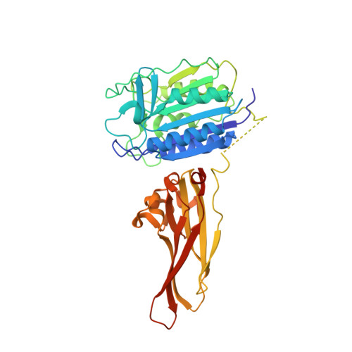
3UOA

Crystal structure of the MALT1 paracaspase (P21 form)


Experimental Data Snapshot

  • Method: X-RAY DIFFRACTION
  • Resolution: 1.75 Å
  • R-Value Free: 0.226 
  • R-Value Work: 0.194 
  • R-Value Observed: 0.196 

wwPDB Validation   3D Report Full Report


This is version 1.4 of the entry. See complete history


Literature

Crystal structure of the mucosa-associated lymphoid tissue lymphoma translocation 1 (MALT1) paracaspase region.

Yu, J.W.Jeffrey, P.D.Ha, J.Y.Yang, X.Shi, Y.

(2011) Proc Natl Acad Sci U S A 108: 21004-21009

  • DOI: https://doi.org/10.1073/pnas.1111708108
  • Primary Citation of Related Structures:  
    3UO8, 3UOA

  • PubMed Abstract: 

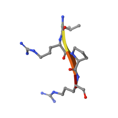
    The mucosa-associated lymphoid tissue lymphoma translocation 1 (MALT1) paracaspase, a key component of the Carma1/Bcl10/MALT1 signalosome, is critical for NF-κB signaling in multiple contexts. MALT1 is thought to function as a scaffold and protease to promote signaling; however, the biochemical and structural basis of paracaspase action remains largely unknown. Here we report the 1.75-Å resolution crystal structure of the MALT1 paracaspase region, which contains the paracaspase domain and an ensuing Ig-like domain. The paracaspase and the Ig domains appear as a single folding unit and interact with each other through extensive van der Waals contacts and hydrogen bonds. The paracaspase domain adopts a fold that is nearly identical to that of classic caspases and homodimerizes similarly to form an active protease. Unlike caspases, the active and mature form of the paracaspase domain remains a single uncleaved polypeptide and specifically recognizes the bound peptide inhibitor Val-Arg-Pro-Arg. In particular, the carboxyl-terminal amino acid Arg of the inhibitor is coordinated by three highly conserved acidic residues. This structure serves as an important framework for deciphering the function and mechanism of paracaspases exemplified by MALT1.


  • Organizational Affiliation

    Department of Molecular Biology, Lewis Thomas Laboratory, Princeton University, Princeton, NJ 08544, USA. jong.x.yu@gsk.com


Macromolecules
Find similar proteins by:  (by identity cutoff)  |  3D Structure
Entity ID: 1
MoleculeChains Sequence LengthOrganismDetailsImage
Mucosa-associated lymphoid tissue lymphoma translocation protein 1A [auth B],
B [auth C]
390Homo sapiensMutation(s): 0 
Gene Names: MALT1MLT
EC: 3.4.22
UniProt & NIH Common Fund Data Resources
Find proteins for Q9UDY8 (Homo sapiens)
Explore Q9UDY8 
Go to UniProtKB:  Q9UDY8
PHAROS:  Q9UDY8
GTEx:  ENSG00000172175 
Entity Groups  
Sequence Clusters30% Identity50% Identity70% Identity90% Identity95% Identity100% Identity
UniProt GroupQ9UDY8
Sequence Annotations
Expand
  • Reference Sequence

Find similar proteins by:  Sequence   |   3D Structure  

Entity ID: 2
MoleculeChains Sequence LengthOrganismDetailsImage
Z-Val-Arg-Pro-DL-Arg-fluoromethylketoneC [auth L],
D [auth M]
6N/AMutation(s): 0 
Sequence Annotations
Expand
  • Reference Sequence
Experimental Data & Validation

Experimental Data

  • Method: X-RAY DIFFRACTION
  • Resolution: 1.75 Å
  • R-Value Free: 0.226 
  • R-Value Work: 0.194 
  • R-Value Observed: 0.196 
  • Space Group: P 1 21 1
Unit Cell:
Length ( Å )Angle ( ˚ )
a = 63.475α = 90
b = 75.735β = 88.98
c = 87.831γ = 90
Software Package:
Software NamePurpose
SCALEPACKdata scaling
PHENIXrefinement
PDB_EXTRACTdata extraction
CBASSdata collection
DENZOdata reduction
PHASERphasing

Structure Validation

View Full Validation Report


Entry History 

Deposition Data

Revision History  (Full details and data files)

  • Version 1.0: 2011-12-21
    Type: Initial release
  • Version 1.1: 2012-02-15
    Changes: Database references
  • Version 1.2: 2012-12-12
    Changes: Other
  • Version 1.3: 2024-03-20
    Changes: Data collection, Database references, Derived calculations, Refinement description
  • Version 1.4: 2024-10-09
    Changes: Structure summary