3UMR | pdb_00003umr

Crystal structure of the G202D mutant of human G-alpha-i1


Experimental Data Snapshot

  • Method: X-RAY DIFFRACTION
  • Resolution: 2.04 Å
  • R-Value Free: 
    0.210 (Depositor), 0.173 (DCC) 
  • R-Value Work: 
    0.170 (Depositor), 0.165 (DCC) 
  • R-Value Observed: 
    0.172 (Depositor) 

Starting Model: experimental
View more details

wwPDB Validation 3D Report Full Report

Validation slider image for 3UMR

Ligand Structure Quality Assessment 


This is version 1.1 of the entry. See complete history

Literature

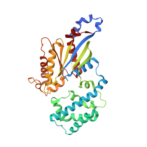
Structural Determinants Underlying the Temperature-sensitive Nature of a G-alpha Mutant in Asymmetric Cell Division of Caenorhabditis elegans

Johnston, C.A.Afshar, K.Snyder, J.T.Tall, G.G.Gonczy, P.Siderovski, D.P.Willard, F.S.

To be published.

Macromolecule Content 

  • Total Structure Weight: 41.13 kDa 
  • Atom Count: 2,981 
  • Modeled Residue Count: 331 
  • Deposited Residue Count: 354 
  • Unique protein chains: 1

Macromolecules

Find similar proteins by:|  3D Structure
Entity ID: 1
MoleculeChains  Sequence LengthOrganismDetailsImage
Guanine nucleotide-binding protein G(i) subunit alpha-1354Homo sapiensMutation(s): 1 
Gene Names: GNAI1
EC: 3.6.5
UniProt & NIH Common Fund Data Resources
Find proteins for P63096 (Homo sapiens)
Explore P63096 
Go to UniProtKB:  P63096
PHAROS:  P63096
GTEx:  ENSG00000127955 
Entity Groups
Sequence Clusters30% Identity50% Identity70% Identity90% Identity95% Identity100% Identity
UniProt GroupP63096
Sequence Annotations
Expand
Reference Sequence

Small Molecules

Ligands 4 Unique
IDChains Name / Formula / InChI Key2D Diagram3D Interactions
GDP

Query on GDP


Download:Ideal Coordinates CCD File
B [auth A]GUANOSINE-5'-DIPHOSPHATE
C10 H15 N5 O11 P2
QGWNDRXFNXRZMB-UUOKFMHZSA-N
SO4

Query on SO4


Download:Ideal Coordinates CCD File
C [auth A]SULFATE ION
O4 S
QAOWNCQODCNURD-UHFFFAOYSA-L
GOL

Query on GOL


Download:Ideal Coordinates CCD File
G [auth A]GLYCEROL
C3 H8 O3
PEDCQBHIVMGVHV-UHFFFAOYSA-N
CL

Query on CL


Download:Ideal Coordinates CCD File
D [auth A],
E [auth A],
F [auth A]
CHLORIDE ION
Cl
VEXZGXHMUGYJMC-UHFFFAOYSA-M

Experimental Data & Validation

Experimental Data

  • Method: X-RAY DIFFRACTION
  • Resolution: 2.04 Å
  • R-Value Free:  0.210 (Depositor), 0.173 (DCC) 
  • R-Value Work:  0.170 (Depositor), 0.165 (DCC) 
  • R-Value Observed: 0.172 (Depositor) 
Space Group: I 4
Unit Cell:
Length ( Å )Angle ( ˚ )
a = 121.952α = 90
b = 121.952β = 90
c = 67.382γ = 90
Software Package:
Software NamePurpose
CrystalCleardata collection
PHASERphasing
PHENIXrefinement
HKL-2000data reduction
HKL-2000data scaling

Structure Validation

View Full Validation Report


Ligand Structure Quality Assessment 


Entry History 

Deposition Data

Revision History  (Full details and data files)

  • Version 1.0: 2012-10-24
    Type: Initial release
  • Version 1.1: 2023-09-13
    Changes: Data collection, Database references, Derived calculations, Refinement description