3ULV | pdb_00003ulv

Structure of quaternary complex of human TLR3ecd with three Fabs (Form2)


Experimental Data Snapshot

  • Method: X-RAY DIFFRACTION
  • Resolution: 3.52 Å
  • R-Value Free: 
    0.253 (Depositor), 0.250 (DCC) 
  • R-Value Work: 
    0.231 (Depositor), 0.229 (DCC) 
  • R-Value Observed: 
    0.232 (Depositor) 

Starting Models: experimental
View more details

wwPDB Validation 3D Report Full Report

Validation slider image for 3ULV

Ligand Structure Quality Assessment 


This is version 2.2 of the entry. See complete history

Literature

Lateral Clustering of TLR3:dsRNA Signaling Units Revealed by TLR3ecd:3Fabs Quaternary Structure.

Luo, J.Obmolova, G.Malia, T.J.Wu, S.J.Duffy, K.E.Marion, J.D.Bell, J.K.Ge, P.Zhou, Z.H.Teplyakov, A.Zhao, Y.Lamb, R.J.Jordan, J.L.San Mateo, L.R.Sweet, R.W.Gilliland, G.L.

(2012) J Mol Biology 421: 112-124

  • DOI: https://doi.org/10.1016/j.jmb.2012.05.006
  • Primary Citation Related Structures: 
    3ULS, 3ULU, 3ULV

  • PubMed Abstract: 

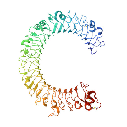
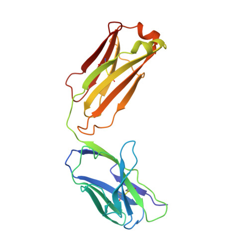
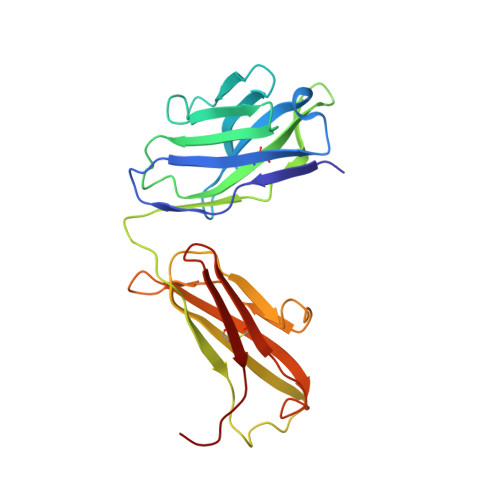
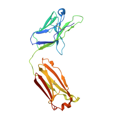
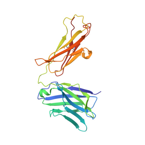
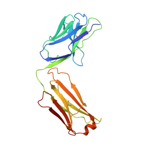
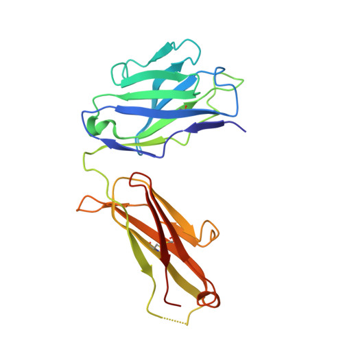
    Toll-like receptor 3 (TLR3) recognizes dsRNA and initiates an innate immune response through the formation of a signaling unit (SU) composed of one double-stranded RNA (dsRNA) and two TLR3 molecules. We report the crystal structure of human TLR3 ectodomain (TLR3ecd) in a quaternary complex with three neutralizing Fab fragments. Fab15 binds an epitope that overlaps the C-terminal dsRNA binding site and, in biochemical assays, blocks the interaction of TLR3ecd with dsRNA, thus directly antagonizing TLR3 signaling through inhibition of SU formation. In contrast, Fab12 and Fab1068 bind TLR3ecd at sites distinct from the N- and C-terminal regions that interact with dsRNA and do not inhibit minimal SU formation with short dsRNA. Molecular modeling based on the co-structure rationalizes these observations by showing that both Fab12 and Fab1068 prevent lateral clustering of SUs along the length of the dsRNA ligand. This model is further supported by cell-based assay results using dsRNA ligands of lengths that support single and multiple SUs. Thus, their antagonism of TLR3 signaling indicates that lateral clustering of SUs is required for TLR3 signal transduction.


  • Organizational Affiliation
    • Biologics Research, Janssen Research and Development, L.L.C., 145 King of Prussia Road, Radnor, PA 19087, USA. jluo@its.jnj.com

Macromolecule Content 

  • Total Structure Weight: 226.08 kDa 
  • Atom Count: 15,591 
  • Modeled Residue Count: 1,973 
  • Deposited Residue Count: 2,010 
  • Unique protein chains: 7

Macromolecules

Find similar proteins by:|  3D Structure
Entity ID: 1
MoleculeChains  Sequence LengthOrganismDetailsImage
Toll-like receptor 3694Homo sapiensMutation(s): 0 
Gene Names: TLR3
UniProt & NIH Common Fund Data Resources
Find proteins for O15455 (Homo sapiens)
Explore O15455 
Go to UniProtKB:  O15455
PHAROS:  O15455
GTEx:  ENSG00000164342 
Entity Groups
Sequence Clusters30% Identity50% Identity70% Identity90% Identity95% Identity100% Identity
UniProt GroupO15455
Glycosylation
Glycosylation Sites: 11Go to GlyGen: O15455-1
Sequence Annotations
Expand
Reference Sequence
Find similar proteins by:|  3D Structure
Entity ID: 2
MoleculeChains  Sequence LengthOrganismDetailsImage
Fab15 light chainB [auth L]214Homo sapiensMutation(s): 0 
Find similar proteins by:|  3D Structure
Entity ID: 3
MoleculeChains  Sequence LengthOrganismDetailsImage
Fab15 heavy chainC [auth H]225Homo sapiensMutation(s): 0 
Find similar proteins by:|  3D Structure
Entity ID: 4
MoleculeChains  Sequence LengthOrganismDetailsImage
Fab12 light chainD [auth C]213Homo sapiensMutation(s): 0 
Find similar proteins by:|  3D Structure
Entity ID: 5
MoleculeChains  Sequence LengthOrganismDetailsImage
Fab12 heavy chainE [auth D]226Homo sapiensMutation(s): 0 
Find similar proteins by:|  3D Structure
Entity ID: 6
MoleculeChains  Sequence LengthOrganismDetailsImage
Fab1068 light chainF [auth E]215Homo sapiensMutation(s): 0 
Find similar proteins by:|  3D Structure
Entity ID: 7
MoleculeChains  Sequence LengthOrganismDetailsImage
Fab1068 heavy chainG [auth F]223Homo sapiensMutation(s): 0 

Oligosaccharides

Help  
Entity ID: 8
MoleculeChains Length2D Diagram GlycosylationD Interactions
2-acetamido-2-deoxy-beta-D-glucopyranose-(1-4)-2-acetamido-2-deoxy-beta-D-glucopyranoseH [auth B],
I [auth G],
J [auth I],
L [auth K]
2N-Glycosylation
Glycosylation Resources
GlyTouCan: G42666HT
GlyCosmos: G42666HT
GlyGen: G42666HT
Entity ID: 9
MoleculeChains Length2D Diagram GlycosylationD Interactions
alpha-D-mannopyranose-(1-6)-beta-D-mannopyranose-(1-4)-2-acetamido-2-deoxy-beta-D-glucopyranose-(1-4)-2-acetamido-2-deoxy-beta-D-glucopyranoseK [auth J]4N-Glycosylation
Glycosylation Resources
GlyTouCan: G22573RC
GlyCosmos: G22573RC
GlyGen: G22573RC

Small Molecules

Ligands 2 Unique
IDChains Name / Formula / InChI Key2D Diagram3D Interactions
NAG

Query on NAG


Download:Ideal Coordinates CCD File
M [auth A]
N [auth A]
O [auth A]
P [auth A]
Q [auth A]
M [auth A],
N [auth A],
O [auth A],
P [auth A],
Q [auth A],
R [auth A]
2-acetamido-2-deoxy-beta-D-glucopyranose
C8 H15 N O6
OVRNDRQMDRJTHS-FMDGEEDCSA-N
SO4

Query on SO4


Download:Ideal Coordinates CCD File
S [auth A]
T [auth A]
U [auth A]
V [auth A]
W [auth E]
S [auth A],
T [auth A],
U [auth A],
V [auth A],
W [auth E],
X [auth E]
SULFATE ION
O4 S
QAOWNCQODCNURD-UHFFFAOYSA-L

Experimental Data & Validation

Experimental Data

  • Method: X-RAY DIFFRACTION
  • Resolution: 3.52 Å
  • R-Value Free:  0.253 (Depositor), 0.250 (DCC) 
  • R-Value Work:  0.231 (Depositor), 0.229 (DCC) 
  • R-Value Observed: 0.232 (Depositor) 
Space Group: C 1 2 1
Unit Cell:
Length ( Å )Angle ( ˚ )
a = 363.3α = 90
b = 131.66β = 91.35
c = 154.2γ = 90
Software Package:
Software NamePurpose
StructureStudiodata collection
PHASERphasing
PHENIXrefinement
XDSdata reduction
XDSdata scaling

Structure Validation

View Full Validation Report


Ligand Structure Quality Assessment 


Entry History 

Deposition Data

Revision History  (Full details and data files)

  • Version 1.0: 2012-05-23
    Type: Initial release
  • Version 1.1: 2012-05-30
    Changes: Database references
  • Version 1.2: 2012-07-25
    Changes: Database references
  • Version 2.0: 2020-07-29
    Type: Remediation
    Reason: Carbohydrate remediation
    Changes: Atomic model, Data collection, Database references, Derived calculations, Structure summary
  • Version 2.1: 2023-09-13
    Changes: Data collection, Database references, Refinement description, Structure summary
  • Version 2.2: 2024-11-20
    Changes: Structure summary