3UFK | pdb_00003ufk

Crystal structure of UndA complexed with Iron Nitrilotriacetate


Experimental Data Snapshot

  • Method: X-RAY DIFFRACTION
  • Resolution: 2.10 Å
  • R-Value Free: 
    0.205 (Depositor), 0.204 (DCC) 
  • R-Value Work: 
    0.155 (Depositor), 0.156 (DCC) 
  • R-Value Observed: 
    0.158 (Depositor) 

wwPDB Validation 3D Report Full Report

Validation slider image for 3UFK

Ligand Structure Quality Assessment 


This is version 2.1 of the entry. See complete history

Literature

The Crystal Structure of the Extracellular 11-heme Cytochrome UndA Reveals a Conserved 10-heme Motif and Defined Binding Site for Soluble Iron Chelates.

Edwards, M.J.Hall, A.Shi, L.Fredrickson, J.K.Zachara, J.M.Butt, J.N.Richardson, D.J.Clarke, T.A.

(2012) Structure 20: 1275-1284

  • DOI: https://doi.org/10.1016/j.str.2012.04.016
  • Primary Citation Related Structures: 
    3UCP, 3UFH, 3UFK

  • PubMed Abstract: 

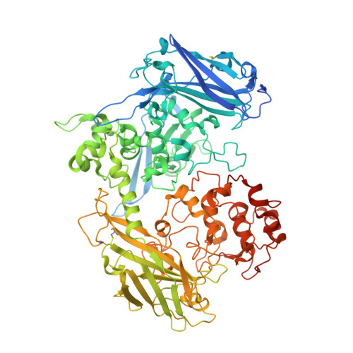
    Members of the genus Shewanella translocate deca- or undeca-heme cytochromes to the external cell surface thus enabling respiration using extracellular minerals and polynuclear Fe(III) chelates. The high resolution structure of the first undeca-heme outer membrane cytochrome, UndA, reveals a crossed heme chain with four potential electron ingress/egress sites arranged within four domains. Sequence and structural alignment of UndA and the deca-heme MtrF reveals the extra heme of UndA is inserted between MtrF hemes 6 and 7. The remaining UndA hemes can be superposed over the heme chain of the decaheme MtrF, suggesting that a ten heme core is conserved between outer membrane cytochromes. The UndA structure has also been crystallographically resolved in complex with substrates, an Fe(III)-nitrilotriacetate dimer or an Fe(III)-citrate trimer. The structural resolution of these UndA-Fe(III)-chelate complexes provides a rationale for previous kinetic measurements on UndA and other outer membrane cytochromes.


  • Organizational Affiliation
    • Centre for Molecular and Structural Biochemistry, School of Biological Sciences and School of Chemistry, University of East Anglia, Norwich Research Park, Norwich NR4 7TJ, UK.

Macromolecule Content 

  • Total Structure Weight: 100.77 kDa 
  • Atom Count: 7,516 
  • Modeled Residue Count: 800 
  • Deposited Residue Count: 874 
  • Unique protein chains: 1

Macromolecules

Find similar proteins by:|  3D Structure
Entity ID: 1
MoleculeChains  Sequence LengthOrganismDetailsImage
UndA874Shewanella sp. HRCR_06Mutation(s): 0 
Gene Names: unda
UniProt
Find proteins for F8UWD6 (Shewanella sp. HRCR_06)
Explore F8UWD6 
Go to UniProtKB:  F8UWD6
Entity Groups
Sequence Clusters30% Identity50% Identity70% Identity90% Identity95% Identity100% Identity
UniProt GroupF8UWD6
Sequence Annotations
Expand
Reference Sequence

Small Molecules

Ligands 7 Unique
IDChains Name / Formula / InChI Key2D Diagram3D Interactions
HEC

Query on HEC


Download:Ideal Coordinates CCD File
B [auth A]
C [auth A]
D [auth A]
E [auth A]
F [auth A]
B [auth A],
C [auth A],
D [auth A],
E [auth A],
F [auth A],
G [auth A],
H [auth A],
I [auth A],
J [auth A],
K [auth A],
L [auth A]
HEME C
C34 H34 Fe N4 O4
HXQIYSLZKNYNMH-LJNAALQVSA-N
NTA

Query on NTA


Download:Ideal Coordinates CCD File
S [auth A],
T [auth A],
X [auth A],
Y [auth A]
NITRILOTRIACETIC ACID
C6 H9 N O6
MGFYIUFZLHCRTH-UHFFFAOYSA-N
GOL

Query on GOL


Download:Ideal Coordinates CCD File
AA [auth A],
BA [auth A],
CA [auth A],
Z [auth A]
GLYCEROL
C3 H8 O3
PEDCQBHIVMGVHV-UHFFFAOYSA-N
FE

Query on FE


Download:Ideal Coordinates CCD File
P [auth A],
Q [auth A],
U [auth A],
V [auth A]
FE (III) ION
Fe
VTLYFUHAOXGGBS-UHFFFAOYSA-N
CA

Query on CA


Download:Ideal Coordinates CCD File
M [auth A]CALCIUM ION
Ca
BHPQYMZQTOCNFJ-UHFFFAOYSA-N
MG

Query on MG


Download:Ideal Coordinates CCD File
N [auth A],
O [auth A]
MAGNESIUM ION
Mg
JLVVSXFLKOJNIY-UHFFFAOYSA-N
O

Query on O


Download:Ideal Coordinates CCD File
R [auth A],
W [auth A]
OXYGEN ATOM
O
XLYOFNOQVPJJNP-UHFFFAOYSA-N

Experimental Data & Validation

Experimental Data

  • Method: X-RAY DIFFRACTION
  • Resolution: 2.10 Å
  • R-Value Free:  0.205 (Depositor), 0.204 (DCC) 
  • R-Value Work:  0.155 (Depositor), 0.156 (DCC) 
  • R-Value Observed: 0.158 (Depositor) 
Space Group: P 21 21 21
Unit Cell:
Length ( Å )Angle ( ˚ )
a = 69.5α = 90
b = 105.87β = 90
c = 151.52γ = 90
Software Package:
Software NamePurpose
xia2data scaling
PHASERphasing
REFMACrefinement
xia2data reduction

Structure Validation

View Full Validation Report


Ligand Structure Quality Assessment 


Entry History 

Deposition Data

Revision History  (Full details and data files)

  • Version 1.0: 2012-08-08
    Type: Initial release
  • Version 2.0: 2023-11-15
    Changes: Atomic model, Data collection, Database references, Derived calculations
  • Version 2.1: 2024-11-20
    Changes: Structure summary